Malignant neuroendocrine tumor with epithelial differentiation
Findings:
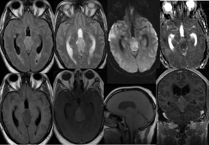
Multiple MRI images demonstrate a heterogeneously enhancing mass in the region of the pineal gland. The lesion demonstrates intermediate T2 signal and mild restricted diffusion indicating a cellular lesion. The mass splays the third ventricle posterior aspect and compresses the tectum, causing obstructive hydrocephalus with a small amount of transependymal CSF flow. A small indeterminate FLAIR hyperintense nodule is present within the right temporal horn. The mass has multilobulated margins.
Discussion/Differential Diagnosis:
BACK TO
MAIN PAGE