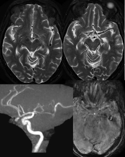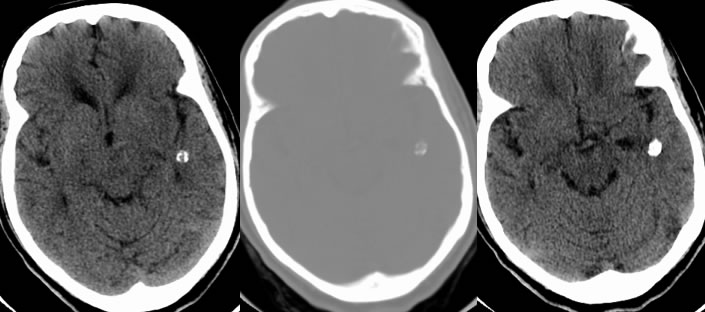

Calcified Left MCA aneurysm
Findings:
Multiple axial CT images demonstrate a partially calcified lesion projected over the left temporal lobe. There is frontal periventricular low attenuation due to chronic microvascular ischemic disease. The MRA images demonstrate contiguity of the calcified lesion with the left MCA posterior division.
Discussion/Differential Diagnosis:
BACK TO
MAIN PAGE

