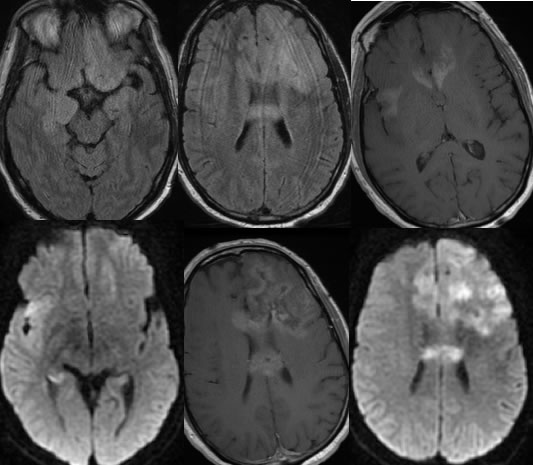
10 months later
Gliomatosis Cerebri, progressive
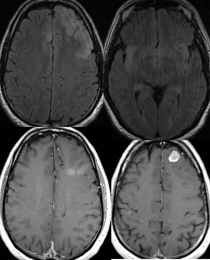
Findings:
Multiple axial MRI images demonstrate poorly defined infiltrative signal abnormality within the left frontal lobe which is associated with several nodular zones of enhancement. Additional infiltrative signal abnormality involves the right frontal parasagittal region and bilateral insular cortex.
The follow up MRI images demonstrate progressive signal abnormalities and enhancement, with new enhancing masses involving the corpus callosum and development of enhancement within the right insular lesion. The corpus callosum mass demonstrates restricted diffusion. Patchy diffusion hyperintensities are also present in the bilateral frontal lobes left greater than right.
Discussion/Differential Diagnosis:
BACK TO
MAIN PAGE