Glioblastoma Multiforme
Findings:
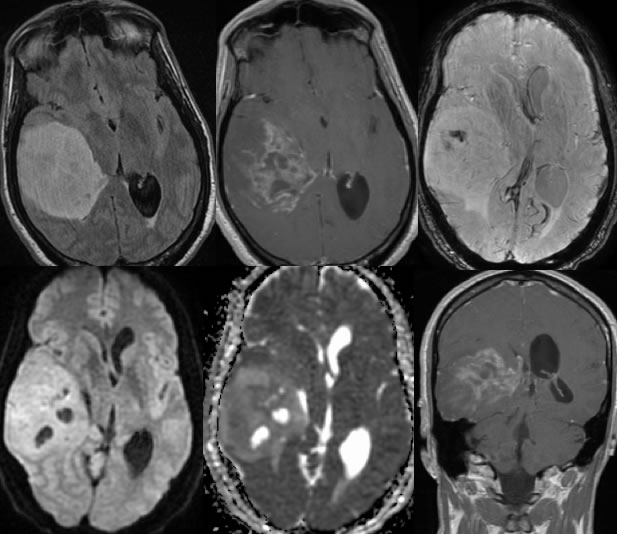
Axial FLAIR image demonstrates a well circumscribed heterogeneously hyperintense lesion in the right temporal lobe. The post contrast images demonstrate heterogeneous enhancement of the lesion with a few zones of cystic change and/or necrosis. There is associated mass effect and midline shift. Mild ventricular trapping is seen on the left. The mass is inseparable from the right lateral ventricle. Susceptibility weighted imaging demonstrates a focus of hemorrhage within the anterior aspect of the mass. Relative diffusion hyperintensity is overall T2 shine through.
Discussion/Differential Diagnosis:
BACK TO
MAIN PAGE