Basilar artery thrombosis
Findings:
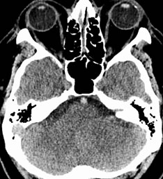
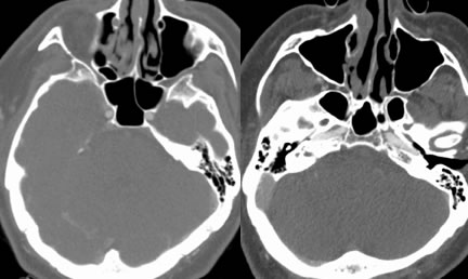
Noncontrast CT image demonstrates asymmetric hyperdensity of the basilar artery. CTA source images demonstrate absent enhancement within this portion of the basilar artery. Coronal and sagittal MPR images demonstrate absent enhancement within the mid portion of the basilar artery.
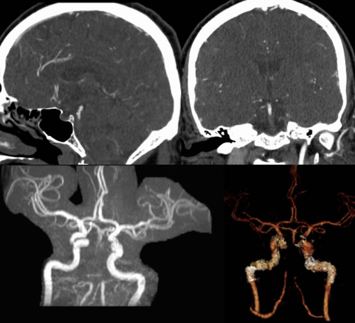
MRA and CTA reformats confirm the presence of occlusive thrombosis of the basilar artery extending from approximately the vertebrobasilar junction through the distal third.
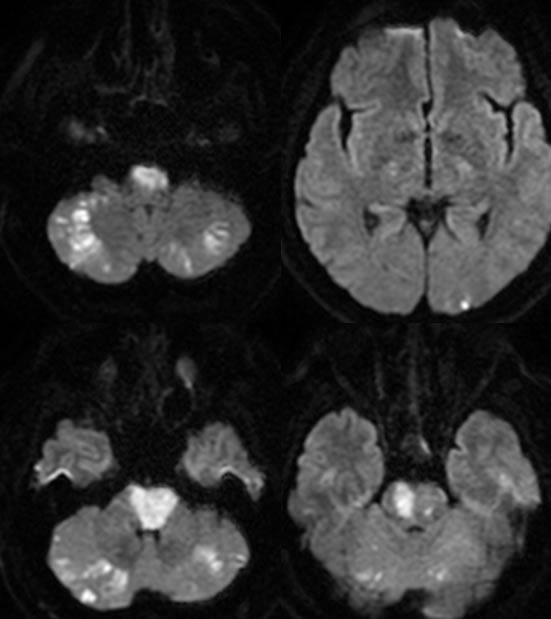
Diffusion weighted images demonstrate multiple acute posterior circulation infarcts, including the bilateral cerebellar hemispheres, pons, and left occipital lobe.
Discussion/Differential Diagnosis:
BACK TO
MAIN PAGE