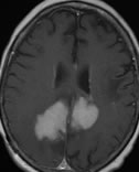
2015
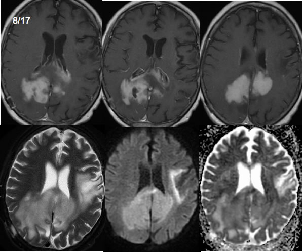
1/2017
Multiple Sclerosis with T cell lymphoma
Findings:
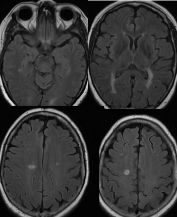
MR images from 2015 demonstrate ovoid periventricular white matter lesions due to multiple sclerosis.
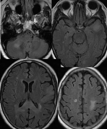
In January 2017, additional confluent signal abnormalities have developed within the left temporal lobe, corpus callosum splenium, and the left parietal subcortical region. Bandlike nearly symmetric enhancement has developed within the corpus callosum splenium anterior aspect, with subtle patchy enhancement in the right centrum semiovale and an ovoid enhancing lesion in the right cerebellar white matter.
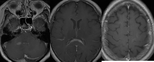
On the March 2017 examination, the bandlike enhancement of the anterior splenium corpus callosum has resolved, but there are now more confluent zones of abnormal enhancement within the superior margin of the corpus callosum splenium and bilateral parietooccipital lobes right greater than left.
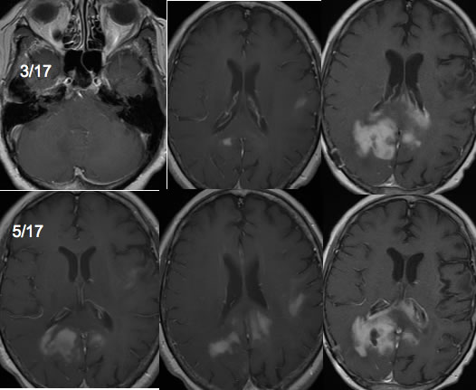
On the May 2017 examination, progressive enhancement is seen with a blotchy distribution, associated with evolving subependymal enhancement within the lateral ventricles. A right parietal burr hole for biopsy is noted with a biopsy cavity within the enhancing mass.
A further slight progression of the abnormal enhancement as seen on the August 2017 examination. The confluent mass demonstrates relative decreased T2 signal and diffusion restriction. Additional confluent zones of diffusion restriction and T2 signal abnormality has developed in the left frontal parietal region
Discussion/Differential Diagnosis:
BACK TO
MAIN PAGE