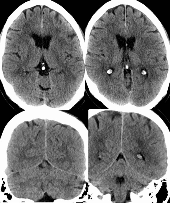
Syncope and fall, exam at 0000. Anything you could call here?
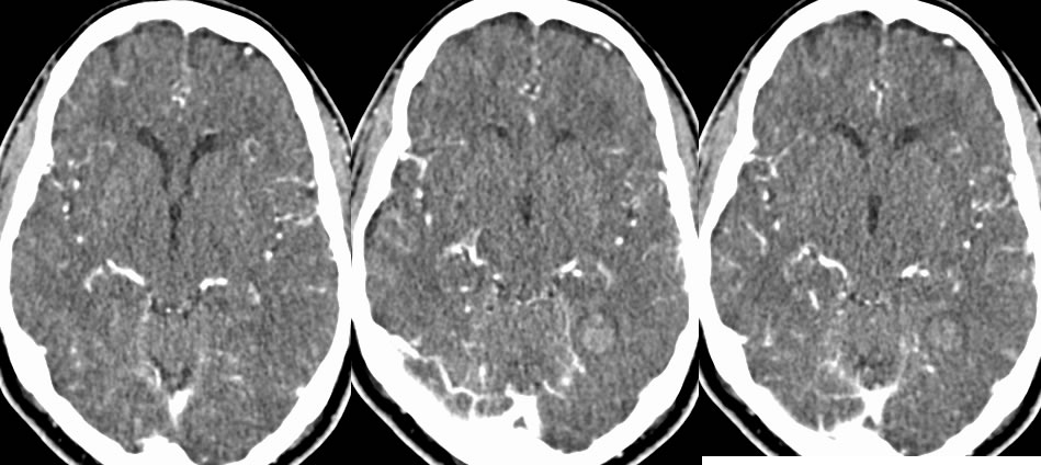
CTA at 0100
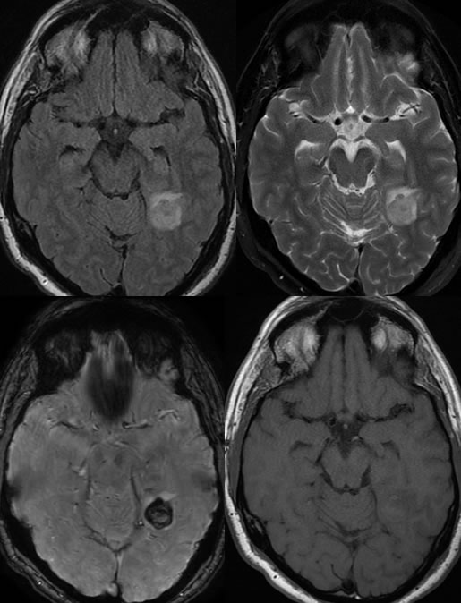
MRI at 0800
Hyperacute Parenchymal Hemorrhage
Findings:
Axial and coronal CT images demonstrate a subtle area of isodensity within the right occipital lobe.
CTA images demonstrate a subtle zone of hyperdensity within the right occipital lobe which was previously isodense.
Follow up MRI images performed eight hours later demonstrate a zone of hemorrhage in the right occipital lobe with surrounding edema. The signal characteristics are compatible with recent hemorrhage.
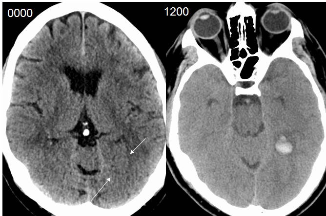
The follow-up noncontrast CT performed 12 hours after the initial noncontrast CT demonstrates a zone of hemorrhage within the right occipital lobe which was isodense 12 hours previously.
Discussion/Differential Diagnosis:
BACK TO
MAIN PAGE