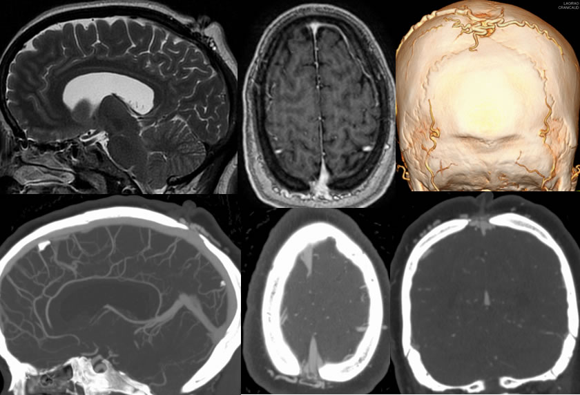
Sinus Pericranii
Findings:
Sagittal T2, axial post contrast T1, coronal VRT images, and MIP CTA images demonstrate a defect in the midline parietal calvarium. Numerous venous structures communicate between the subgaleal scalp and the superior sagittal sinus, extending through the defect.
Discussion/Differential Diagnosis:
BACK TO
MAIN PAGE