Cystic Intradural Lesion
Findings:
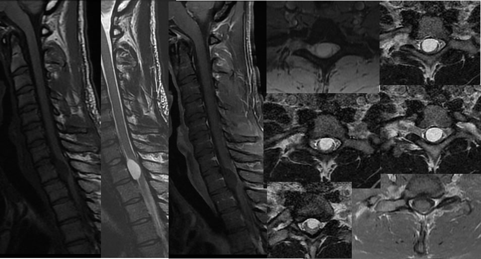
Sagittal T1, sagittal T2, sagittal T1 post contrast images as well as multiple axial images through the cervical spine demonstrate a nonenhancing intradural lesion at the C7 level ventrally, which causes significant displacement and compression of spinal cord parenchyma. On the axial images it is difficult to determine whether this is intramedullary or extra medullary, but there is a relative claw sign of spinal cord parenchyma indicating that it may be intramedullary.
Discussion/Differential Diagnosis:
BACK TO
MAIN PAGE