Fibrous Dysplasia
Findings:
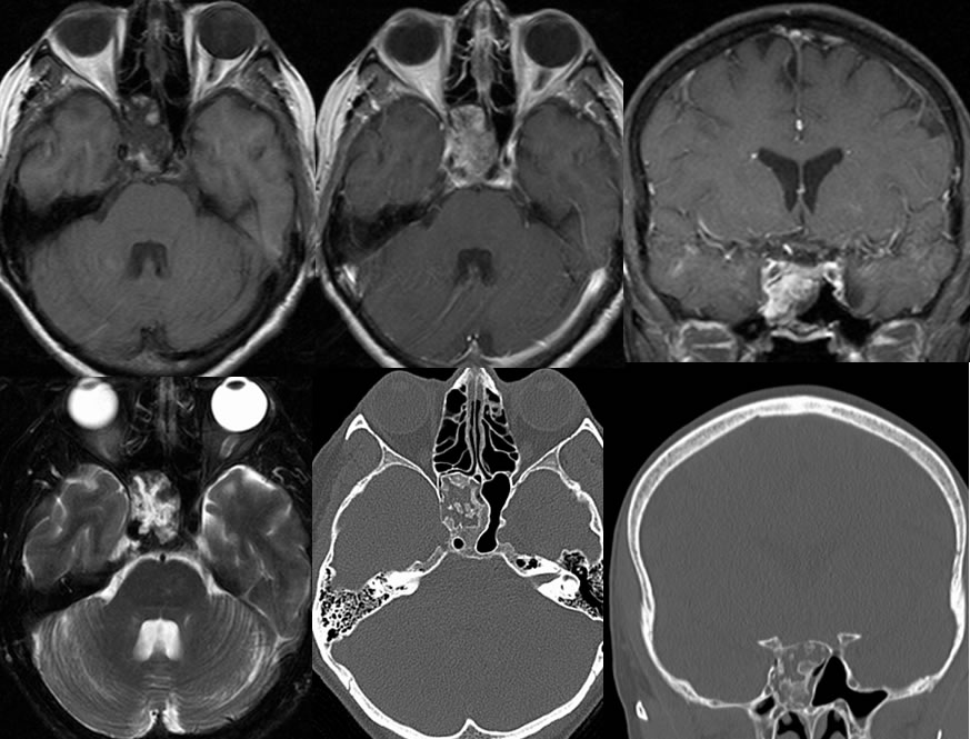
Axial T1, axial T1 post contrast, coronal T1 post contrast with fat saturation, axial T2 with fat saturation, and axial/coronal CT images demonstrate an osseous lesion occupying the right sphenoid sinus. The focus demonstrates heterogeneous enhancement with some zones of fat signal intensity. On the CT images, there is smooth remodeling of the right sphenoid sinus in the region and zones of ground glass bone opacity. The central mildly expansile lytic areas on CT correlate with zones of heterogeneous enhancement after gadolinium administration.
Discussion/Differential Diagnosis:
BACK TO
MAIN PAGE