Florid Osseous Dysplasia and Chronic Osteomyelitis
Findings:
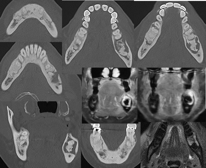
CT and MRI images demonstrate irregular sclerotic lesions within the bilateral mandible, with some lucency around the foci within the left mandibular body having the appearance of central sequestra. A mild expansile appearance is noted. The MR images demonstrate patchy gadolinium enhancement in the region and there is T2 signal alteration surrounding the sclerotic foci in the left mandible. No other abnormal enhancement or T2 hyperintense bone marrow edema signal is identified.
Discussion/Differential Diagnosis:
BACK TO
MAIN PAGE