Fibrous Dysplasia
Findings:
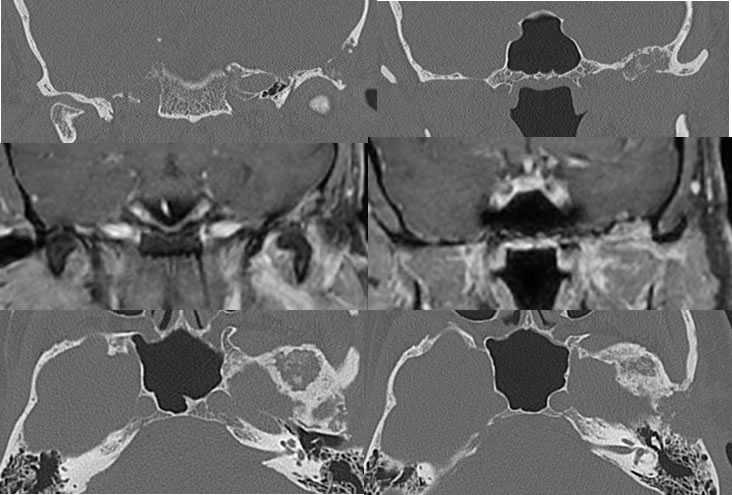
Multiple CT and MRI images demonstrate a mixed lytic and sclerotic process with mild expansile characteristics along the floor of the left middle cranial fossa, with an additional lytic zone along the left condylar fossa. There is mild patchy enhancement in this region on the MRI images.
Discussion/Differential Diagnosis:
BACK TO
MAIN PAGE