Meningitis
Findings:
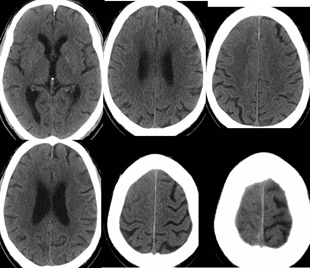
Multiple axial CT images demonstrate asymmetric hyperdensity within right cerebral sulci without significant mass effect. This is most evident near the vertex.
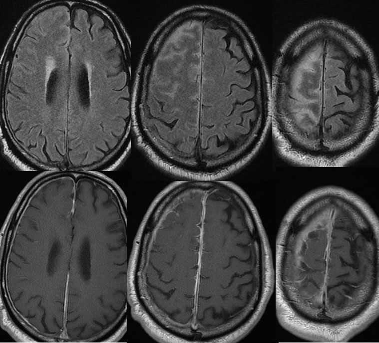
MR images including FLAIR and T1 postcontrast demonstrate abnormal leptomeningeal and pachymeningeal enhancement over the right cerebral convexity, associated with FLAIR hyperintensity of the subarachnoid space.
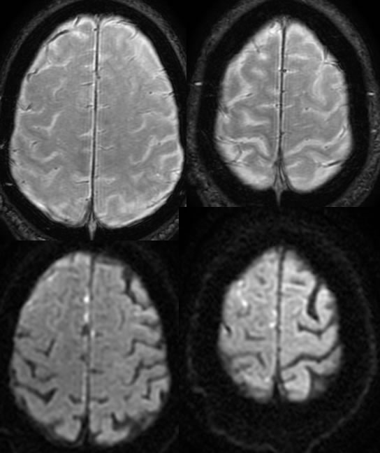
Spotty zones of restricted diffusion are present within this process, with no hemorrhagic staining seen on the SWAN sequence.
Discussion/Differential Diagnosis:
BACK TO
MAIN PAGE