
CNS Vasculitis
Findings:
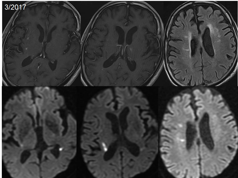
Axial T1 post contrast images and a single diffusion image demonstrate linear foci of abnormal enhancement throughout the deep and subcortical white matter of both frontal and parietal lobes, associated with a few tiny foci of restricted diffusion.
Follow up MRI images two months later demonstrates decreased prominence of the linear foci of enhancement, with development of small foci of enhancement in the right subinsular region and left thalamus. Foci of diffusion restriction are separate from the enhancing lesions. Superimposed ovoid white matter signal alterations are present.
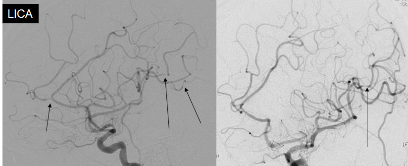
Left internal carotid conventional arteriogram demonstrates multifocal smooth narrowing of anterior and middle cerebral artery branches. Arrows demarcate some but not all foci of vascular narrowing.
Discussion/Differential Diagnosis:
BACK TO
MAIN PAGE