Susac Syndrome, Acute
Findings:
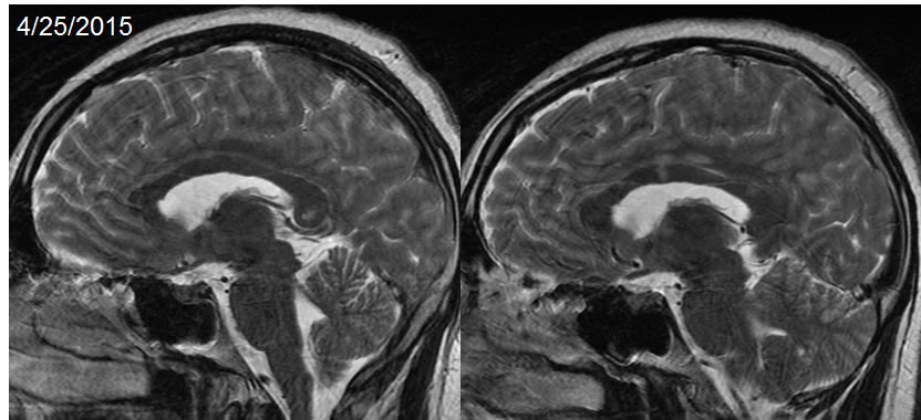
Sagittal T2 weighted MRI from 4/25/2015 demonstrates patchy signal abnormalities within the central portions of the corpus callosum fibers.
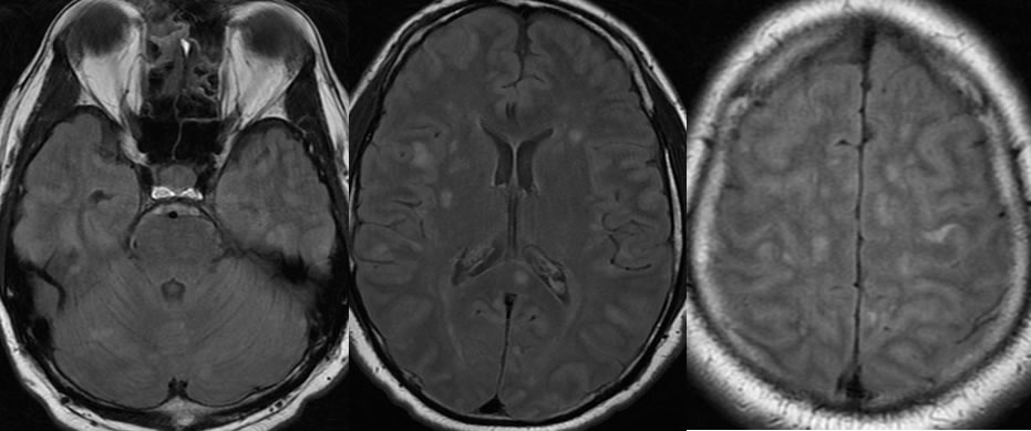
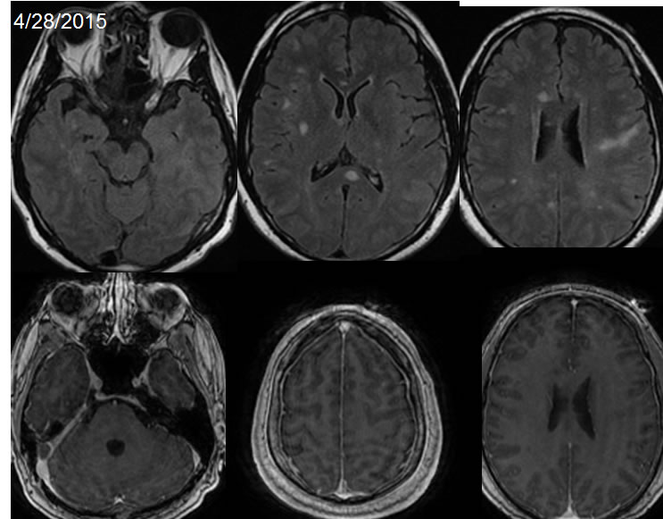
Axial FLAIR images demonstrate multifocal ovoid signal abnormalities within the subcortical and deep white matter of both hemispheres, including the cerebellum and brainstem, with some lesions involving the midportion of the corpus callosum splenium.
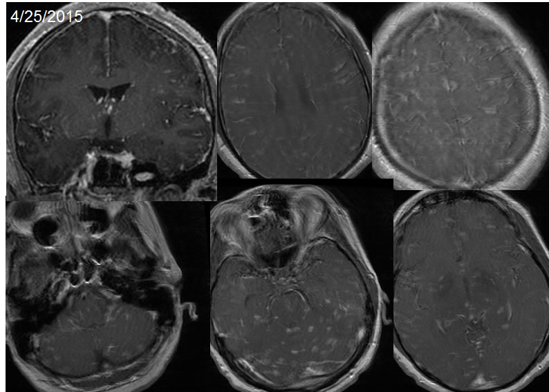
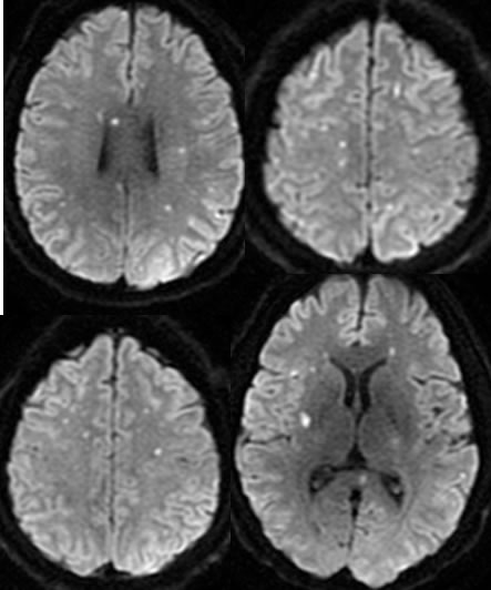
The post contrast T1 weighted imaging dated 4/25/2015 demonstrates extensive ovoid and linear enhancing lesions in both cerebral hemispheres, cerebellum, and brainstem. Multiple foci of restricted diffusion are present. Not all of the enhancing lesions demonstrate diffusion restriction. There is no significant hemorrhagic staining.
The follow up MRI three days later demonstrates essential resolution of the enhancing lesions, with multiple ovoid FLAIR signal alterations including within the central aspect of the corpus callosum.
Discussion/Differential Diagnosis:
BACK TO
MAIN PAGE