Cavernous Sinus Thrombosis
Findings:
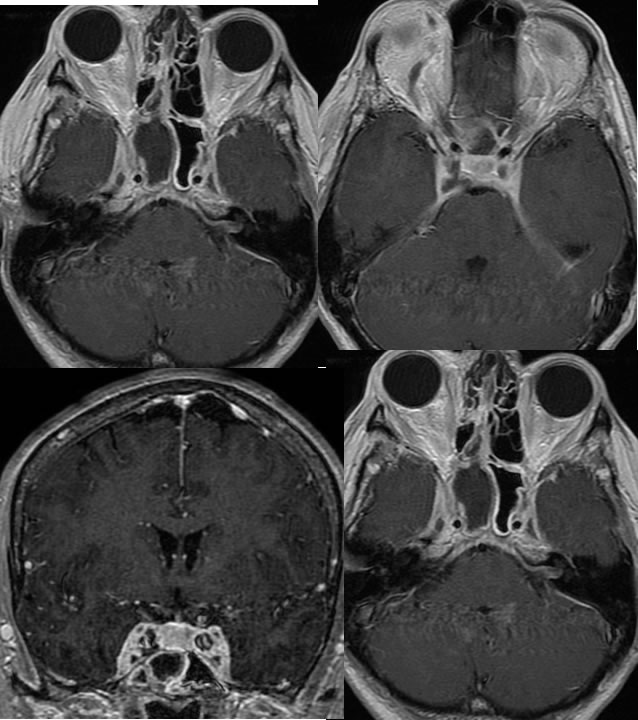
Axial and coronal T1 post contrast images demonstrate extensive fluid signal with surrounding enhancement within the right sphenoid sinus. There is abnormal enhancement within both cavernous sinuses with mild enlargement of the cavernous sinuses. Zones of decreased or absent enhancement are also present within the bilateral cavernous sinuses surrounded by abnormal enhancement.
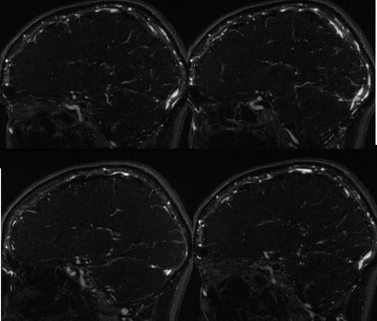
MRI venogram images MIP projection demonstrate absent venous flow signal within the bilateral cavernous sinuses.
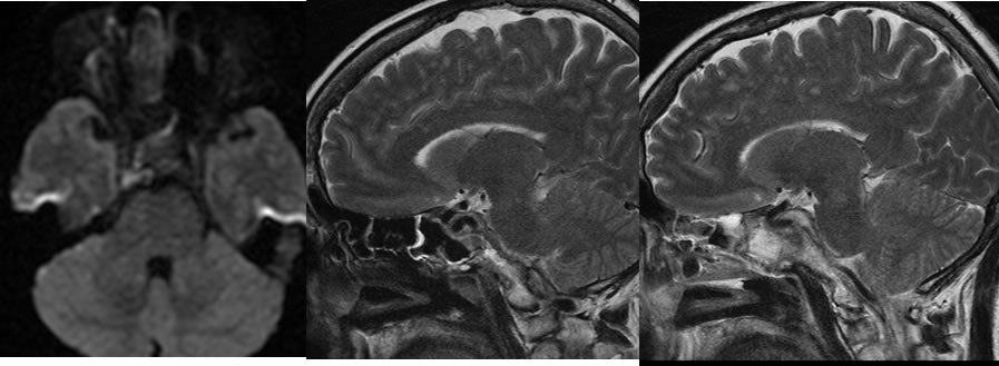
Restricted diffusion is visible within the right cavernous sinus and there is heterogeneous T2 signal abnormality within both cavernous sinuses.
Discussion/Differential Diagnosis:
BACK TO
MAIN PAGE