Tumefactive Multiple Sclerosis
Findings:
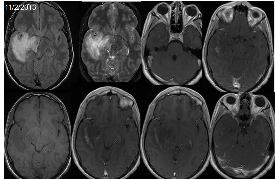
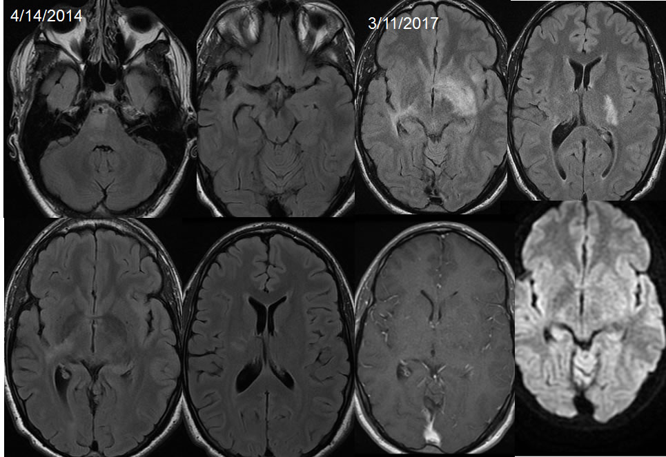
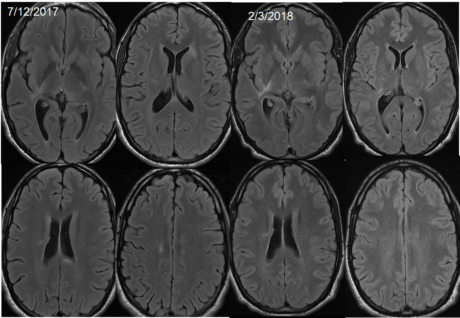
MR images from 2013 demonstrate poorly defined signal abnormality within the right temporal lobe with mass affect and spotty zones of peripheral enhancement. The 2014 images demonstrate near resolution of the process involving the right temporal lobe and external capsule extending into the thalamus. On the 2014 study there is poorly defined signal abnormality of the right pons. On the 2017 study, the left temporal lobe process demonstrates no definite change, with new similar expansile signal alteration within the left basal ganglia that demonstrates no definite diffusion restriction. These processes resolve on 2017 with new signal abnormality in the corpus callosum splenium. The corpus callosum splenium signal abnormality resolves in 2018.
Discussion/Differential Diagnosis:
BACK TO
MAIN PAGE