Hemorrhagic Cord Contusion and R C6 Nerve Root Avulsion
Findings:
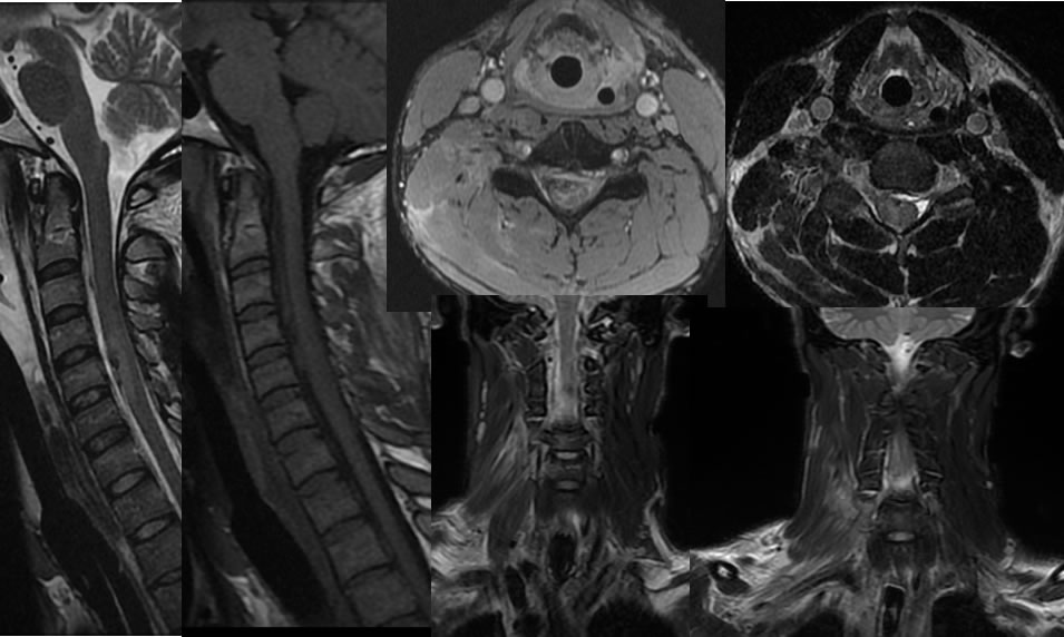
The sagittal T2 weighted image demonstrates normal AP alignment of the cervical spine with mild prevertebral edema signal. There is patchy signal alteration along the ventral spinal cord margin from C4 through C6 with patchy hypointensities ventrally at C5. The sagittal T1 image image demonstrates patchy zones of isointense signal ventral to the mid cervical cord. The axial T2 images demonstrate patchy hemorrhagic changes at the ventral spinal cord margin, including along the right C6 nerve root. Coronal T2 weighted brachial plexus MRI images demonstrate abnormal edema signal within the right scalene musculature extending from C5-6.
Discussion/Differential Diagnosis:
BACK TO
MAIN PAGE