Complex Thoracic Spine Fractures
Findings:
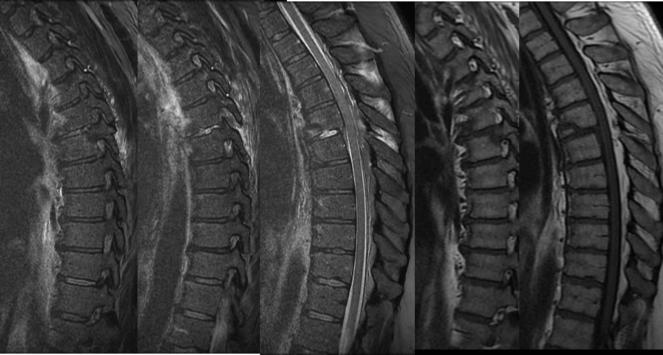
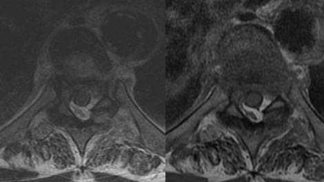
Sagittal MRI images of the thoracic spine demonstrate a complex fracture at the T5 level, which extends through the T5-6 disc space with widening of the disk space and distraction of the T5-6 facet joint, associated with retrolisthesis of T5-6. An additional fracture involves the T6 pedicle and posterior vertebral margin as well as the pars interarticularis seen on the sagittal T-1 sequence. Axial images demonstrate spotty dorsal epidural hematoma and a small ventral epidural hematoma with mild thecal sac narrowing.
Discussion/Differential Diagnosis:
BACK TO
MAIN PAGE