Ankylosing Spondylitis with L2 Posterior Element Fracture
Findings:
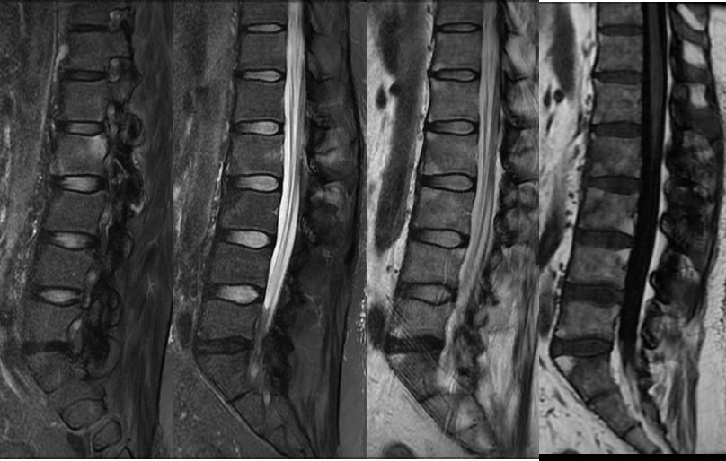
Sagittal MRI images demonstrate straightening and slight kyphosis of the lumbar spine with squared anterior vertebral margins. Foci of fatty marrow conversion involve the corners of several vertebral endplates which have a squared appearance.
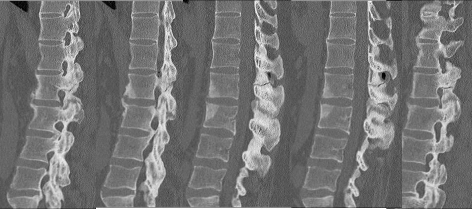
The CT images demonstrate sclerosis at the corners of L2-3 with osteophyte formation. There is anterior longitudinal ligament ossification extending inferiorly to the L1 level. Extensive ankylosis of the bilateral facet joints is present. A nondisplaced fracture extends through posterior elements and spinous process at L2, which also extends through the bilateral pars interarticularis. This is associated with a gas filled chronic synovial cyst like structure.
In retrospect on the MR there is a linear hypointense line extending through the L2 spinous process with marrow edema signal, which correlates with the fracture plane on CT.
Discussion/Differential Diagnosis:
BACK TO
MAIN PAGE