Severe Head Injury with Brain Death CTA
Findings:
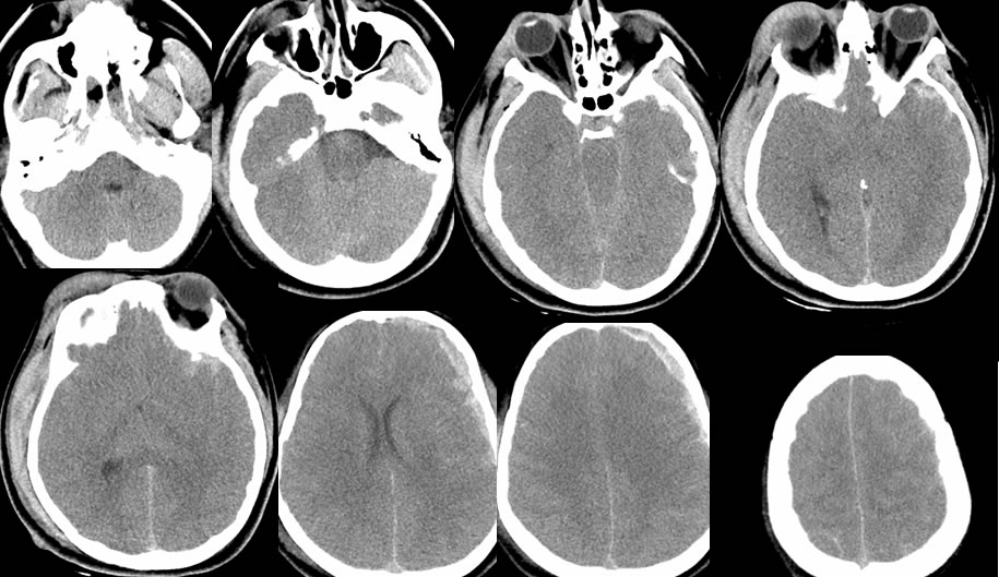
Multiple noncontrast CT images demonstrate extensive traumatic abnormalities, including left convexity subdural hematoma, diffuse spotty subarachnoid hemorrhage, moderate ventricular effacement with cerebral edema, significant deformity of the brainstem due to cerebral edema, with complete effacement of the basal cisterns. There is soft tissue swelling and hematoma over the right orbit and right temporoparietal calvarium with underlying fracture poorly demonstrated.
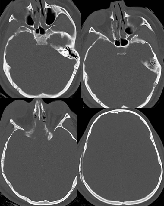
Bone window CT images more optimally demonstrate comminuted displaced right temporal parietal fractures extending through the right sphenoid bone and inferior orbital fissure.
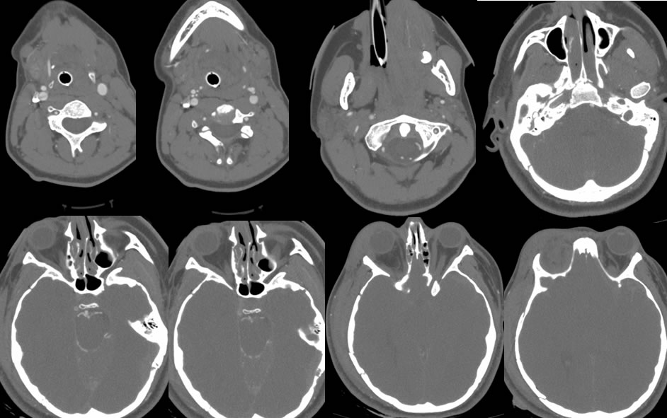
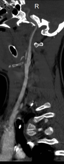
CTA source images demonstrate absent supratentorial flow. There is stagnation of contrast within the basilar artery and proximal aspects of the posterior cerebral arteries. The bilateral carotid bifurcations are patent, but there is no enhancement of the left petrous and cavernous internal carotid and only hazy markedly diminished enhancement of the right cavernous internal carotid.
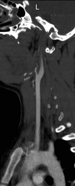
The reformatted images demonstrate what appears to be a tapered occlusion of the left ICA and hazy reduced enhancement of the right ICA distally. This actually represents stagnation of contrast due to markedly increased intracranial pressure exceeding mean arterial pressure.
Discussion/Differential Diagnosis:
BACK TO
MAIN PAGE


