Direct Carotid Cavernous Fistula
Findings:
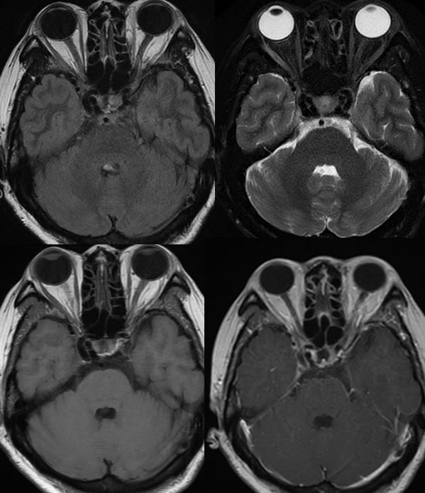
Multiple axial MR images demonstrate abnormal hypointensities within the bilateral cavernous sinuses on the T2 and FLAIR imaging.
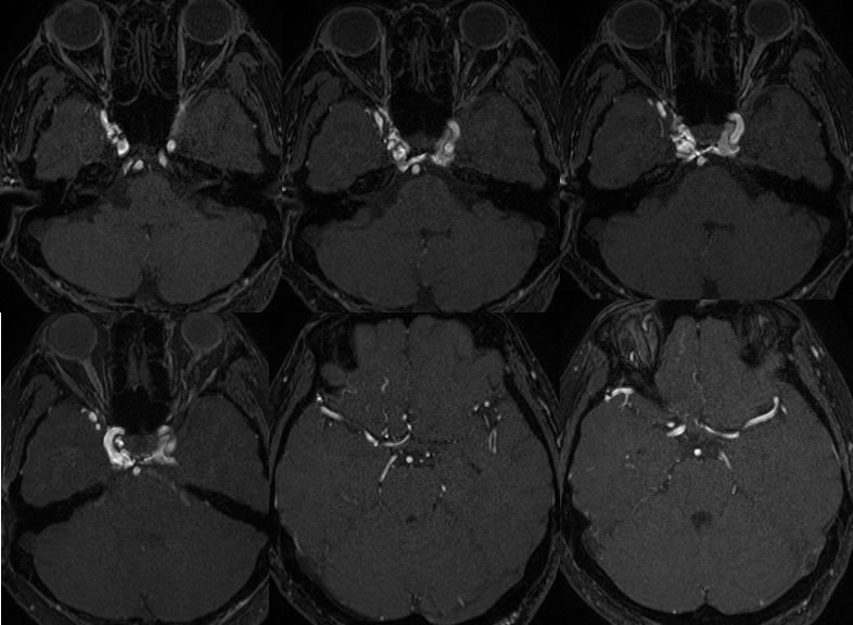
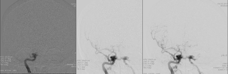
MRA source images demonstrate extensive abnormal arterial signal within the bilateral cavernous sinuses right greater than left. There is also abnormal arterial signal along the right sphenoparietal sinus, inferior right frontal lobe, and minimally involving the right superior ophthalmic vein.
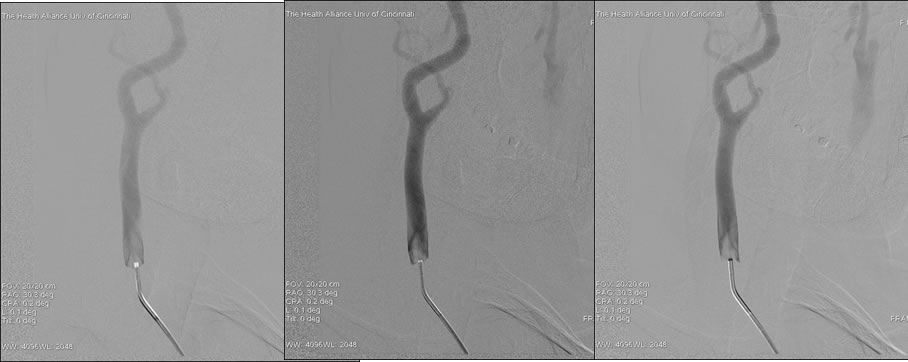
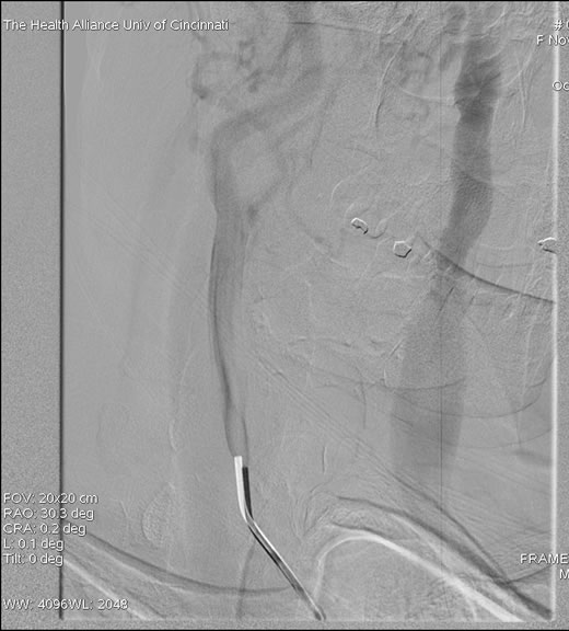
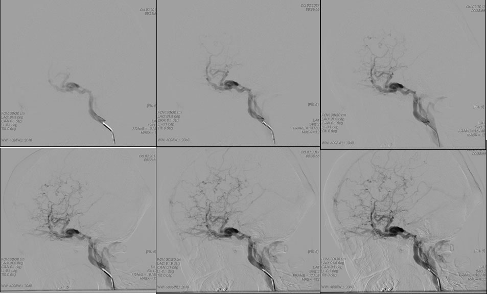
Images from right common carotid arteriogram demonstrate opacification of the left internal jugular vein at the second frame of arterial injection indicating a high flow shunting lesion.
Lateral right internal carotid arteriogram demonstrates immediate cavernous sinus venous opacification at the first frame of injection with extensive retrograde opacification of cortical veins. Shunting into the sphenoparietal and jugular venous systems is seen. On the AP view, the shunting crosses midline to also involve the left cavernous sinus and drains inferiorly through venous plexus.
Discussion/Differential Diagnosis:
BACK TO
MAIN PAGE