Epidural AVM
Findings:
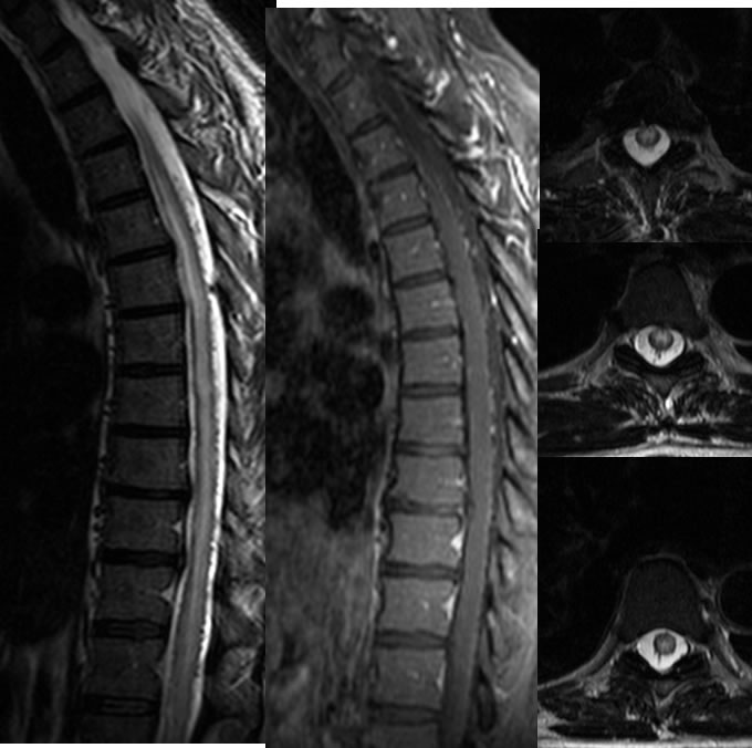
Sagittal T2, sagittal T1 post contrast, and axial T2 weighted images of the thoracic spine demonstrate diffuse thoracic cord edema signal which is predominantly within the central spinal cord parenchyma. Abnormal flow voids with enhancement are present along the dorsal and to a lesser degree ventral aspects of the thoracic cord.
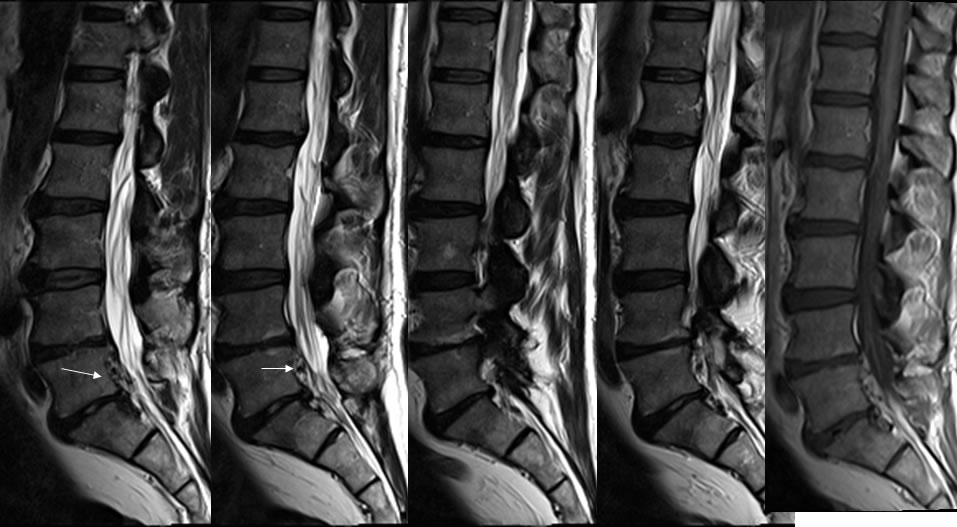
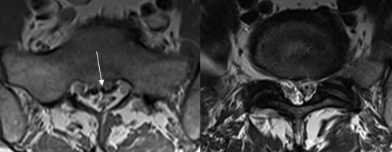
Multiple MR images of the lumbar spine demonstrate multilevel degenerative disc disease. Several spotty and linear T2 hypointensities are present within the ventral epidural space at L5.
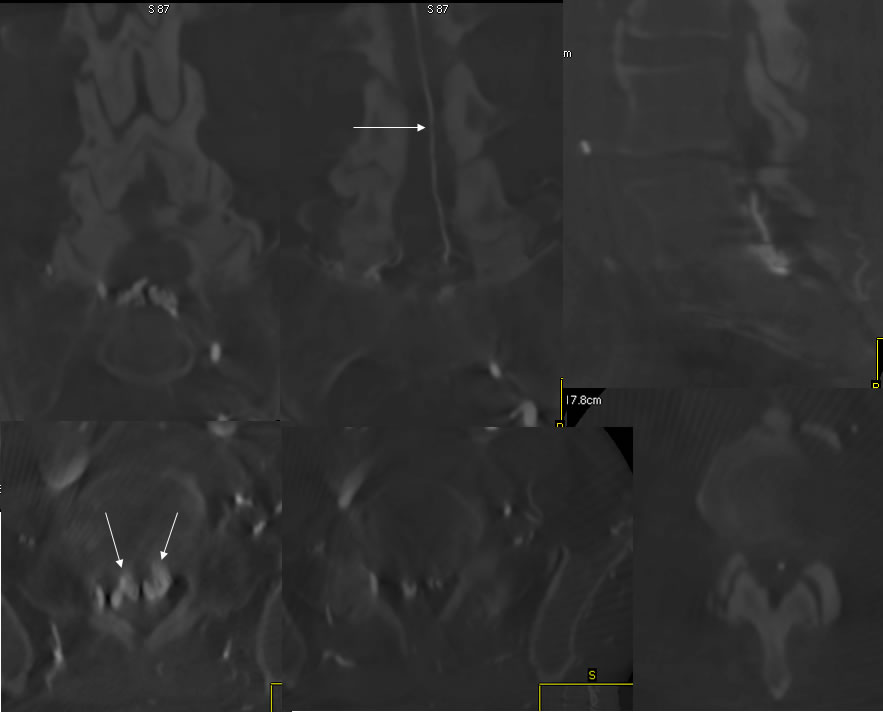
A selective L internal iliac arteriogram with 3D acquisition demonstrates A ventral epidural arteriovenous malformation at the level of L5, associated with a single dominant draining vein extending superiorly toward the thoracic cord.
Discussion/Differential Diagnosis:
BACK TO
MAIN PAGE