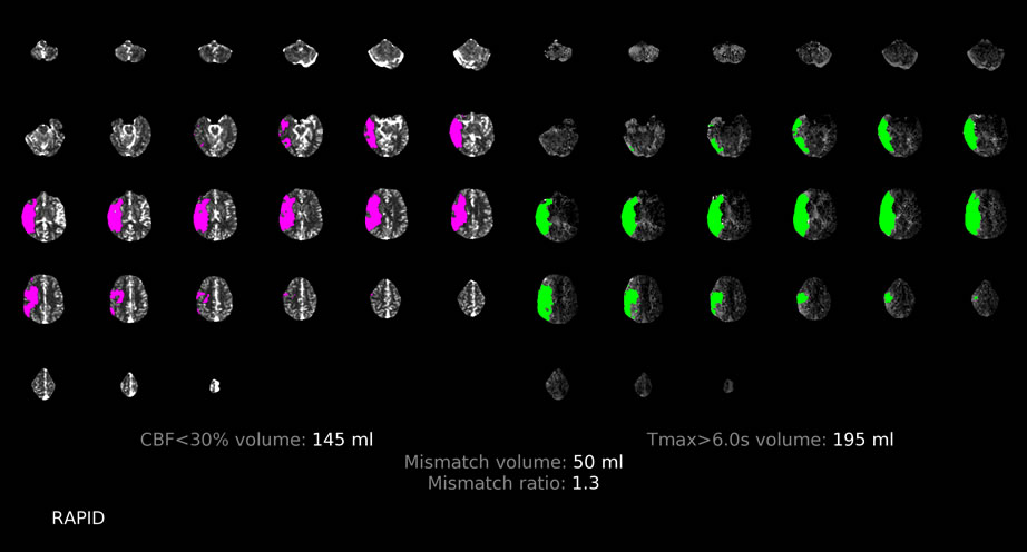
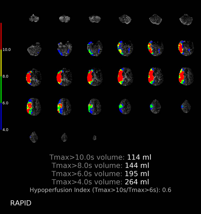
For comparison, please review below, good quality CTP data with nonsalveagable R MCA infarction.
L MCA infarction with invalid perfusion data
Findings:
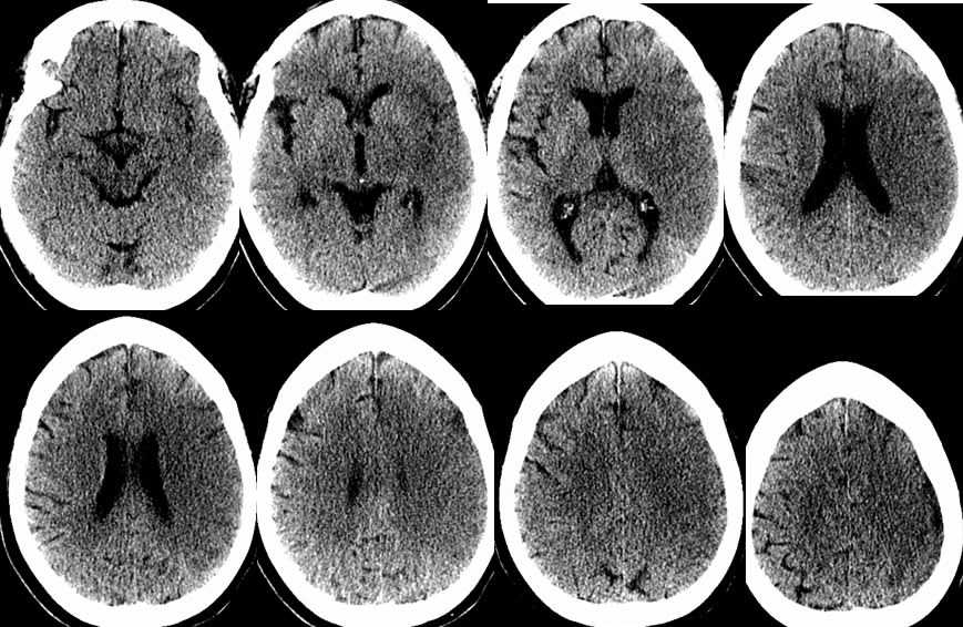
Multiple CT images demonstrate a large wedge shaped zone of low attenuation with sulcal effacement and loss of grey white differentiation involving the left frontotemporal region extending into the basal ganglia and caudate nucleus.
VRT MRA head reformat demonstrates occlusion of the left M1 segment with decreased opacification of distal left MCA branches.
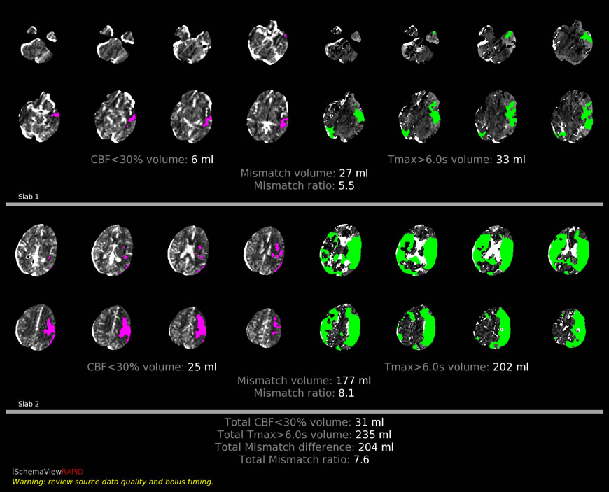
Rapid perfusion study demonstrates large zones of Tmax greater than 6 seconds involving the bilateral cerebral hemispheres, with a moderate zone of hypoperfusion along the left MCA territory. The calculated mismatch ratio is 7.6, which is greater than would be expected for the large well-established left MCA distribution infarct seen on the initial head CT.
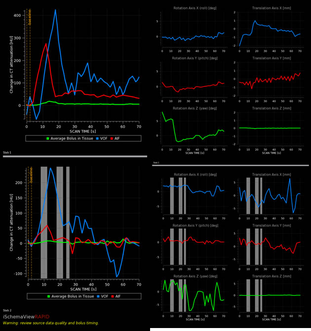
The source data curves demonstrate significant motion artifact, rendering the perfusion data invalid.
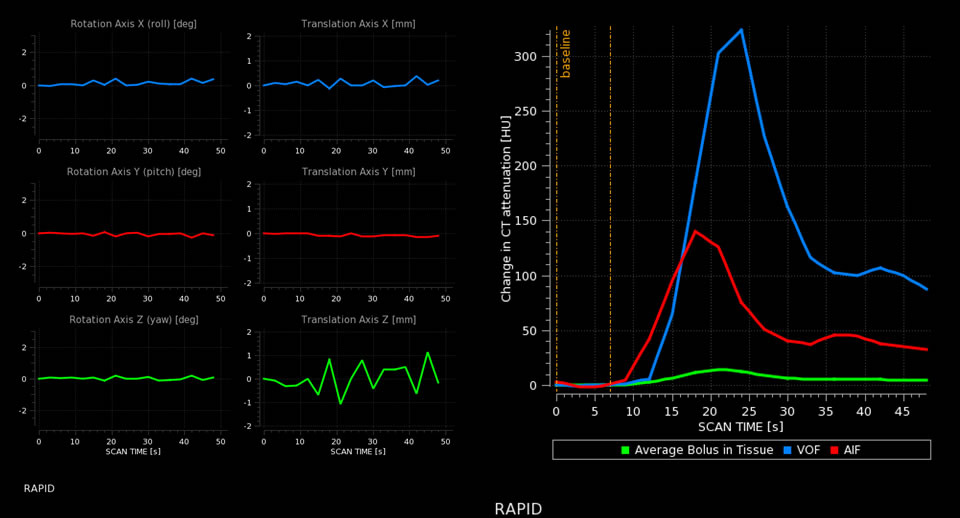
For comparison, better quality data from a different patient is shown, related to a well-established right MCA distribution infarct with no significant salvageable tissue. Some motion is seen on the translation Z axis, but note the smooth VOF and AIF curves compared to the other case.
Discussion/Differential Diagnosis:
BACK TO
MAIN PAGE