R MCA infarction with good quality perfusion data
Findings:
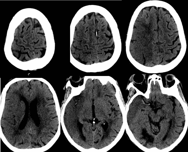
Axial noncontrast CT images demonstrate hyperdensity of the right MCA extending into the bifurcation. There is a large wedge shaped zone of low attenuation involving the right temporal, frontal, and parietal lobes with loss of grey white differentiation and sulcal effacement laterally. The low attenuation extends into the centrum semiovale with some sparing of cortex over the vertex. There is a superimposed remote infarct in the right occipital lobe which is incompletely included. There is superimposed chronic microvascular ischemic white matter disease. Mild mass effect is associated with partial effacement of the right lateral ventricle.
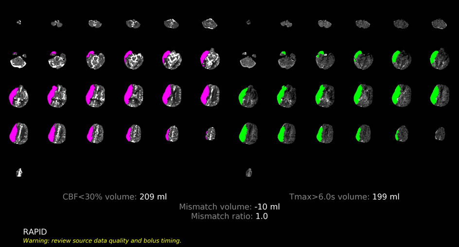
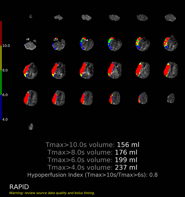
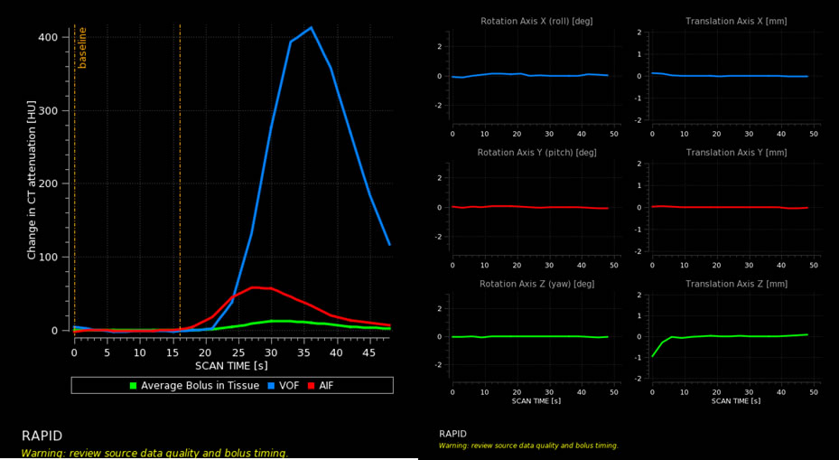
The Rapid perfusion study demonstrates nearly equal volume of markedly reduced blood flow and elevated Tmax, representing a well-established and non-salvageable large MCA distribution infarct. The translation and rotation data is valid, but the imaging should have been continued further since the AIF/VOF curves are truncated.
Discussion/Differential Diagnosis:
BACK TO
MAIN PAGE