Type 2 glomus AVM
Findings:
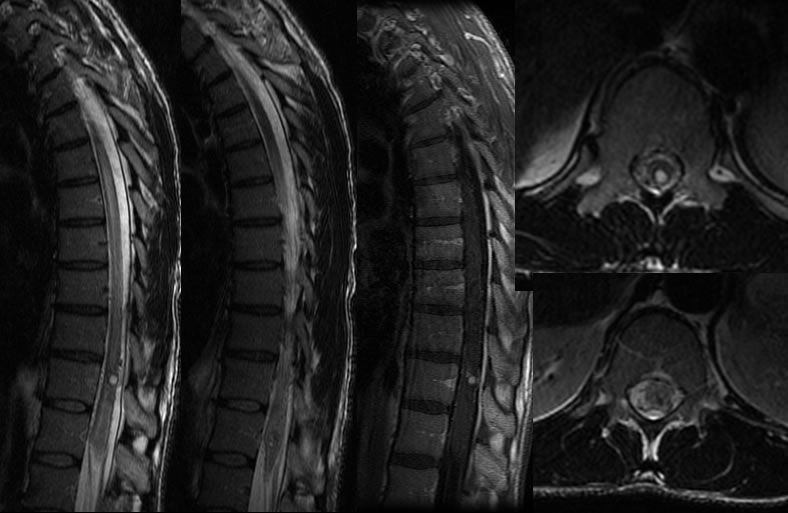
Sagittal T2 weighted images demonstrate patchy T2 hyperintensity within the thoracic cord, associated with a rounded focus of increased T2 signal in the distal thoracic cord. Several abnormal tortuous vessels are seen in the region. The nodular focus demonstrates enhancement after gadolinium administration. The nodular lesion also has a T2 hypointense rim.
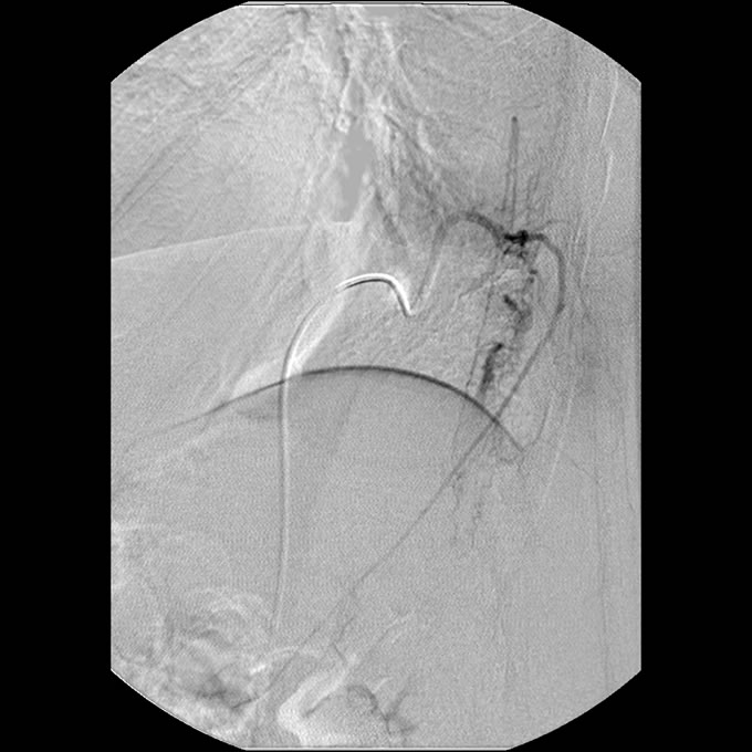
A selective spinal arteriogram demonstrates brisk AV shunting in the region with the nodular focus filling with contrast.
Discussion/Differential Diagnosis:
BACK TO
MAIN PAGE