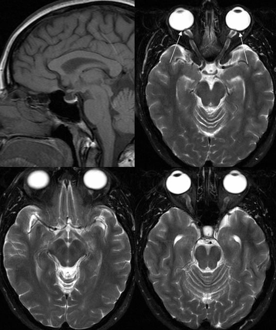
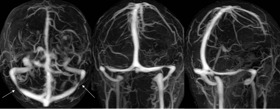
Idiopathic Intracranial Hypertension
Findings:
Mildly flattened pituitary tissue within a borderline size sella is seen on the sagittal T1 image. The axial T2 Image demonstrates flattening of the bilateral posterior globe margins particularly at the optic disc where there is protrusion of tissue. The optic nerve sheaths are dilated and tortuous. MRV images demonstrate symmetric narrowing of the distal transverse sinuses proximal to the transverse sigmoid junctions.
Discussion/Differential Diagnosis:
BACK TO
MAIN PAGE