Hypertrophic olivary degeneration, pontine hemorrhage
Findings:
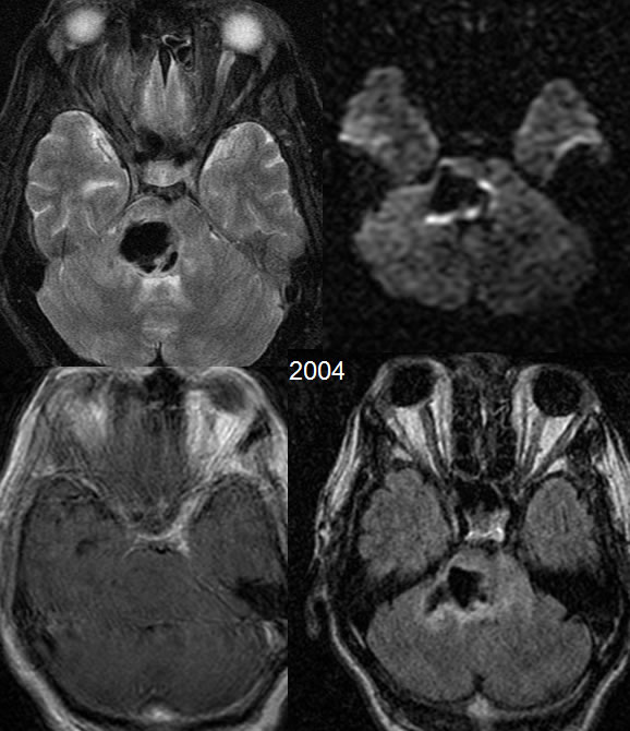
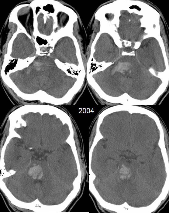
Initial CT and MR images from 2004 demonstrate an acute parenchymal hematoma within the right pons extending into the middle cerebral peduncle. Mild edema is noted. There is mild effacement of the fourth ventricle.
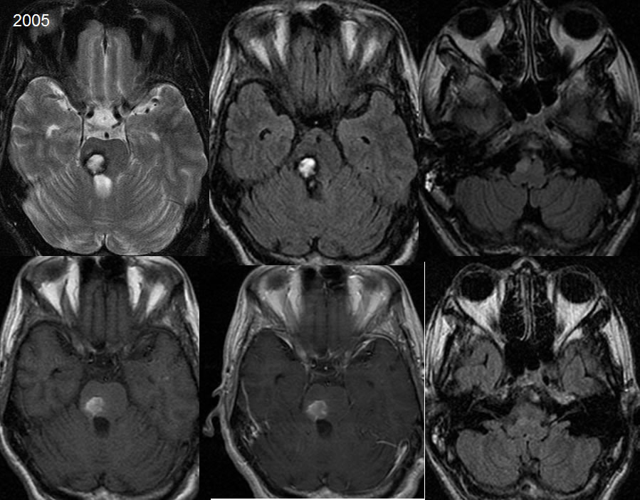
On the 2005 images, the hematoma has contracted and shows expected evolution of blood signal characteristics, with an evolving hemosiderin rim and central zone of extracellular methemoglobin demonstrating hyperintensity on T1 and T2 weighted imaging. The ipsilateral olivary nucleus now demonstrates patchy FLAIR hyperintensity.
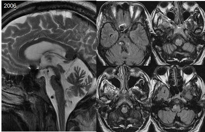
On the 2006 images, there is further contraction of the hematoma with development of brainstem atrophy. The signal abnormality of the right ventral olivary nucleus persists.
Discussion/Differential Diagnosis:
BACK TO
MAIN PAGE