Otosclerosis with obliterative disease
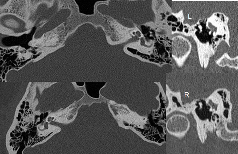
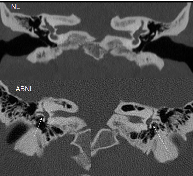
Findings:
Multiple temporal bone CT images demonstrate demineralization surrounding the bilateral cochlea and the anterior oval window margins. Poschl reconstructions show osseous obliteration of both oval windows. The obliterated all windows are also seen on coronal imaging, in comparison to normal aerated oval windows.
Discussion/Differential Diagnosis:
BACK TO
MAIN PAGE