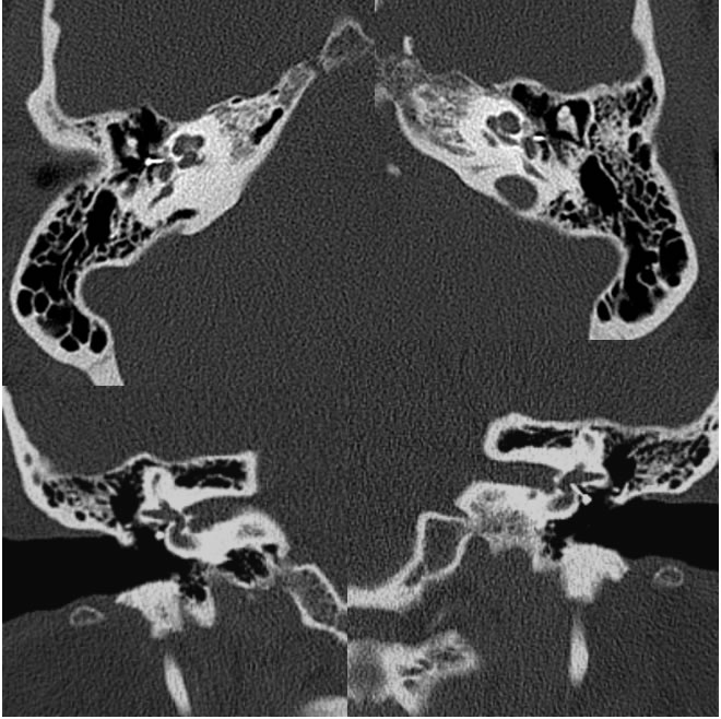
Otosclerosis with stapes prostheses
Findings:
Selected axial and coronal temporal bone CT images demonstrate symmetric demineralization at the bilateral anterior oval window margins. Metallic piston type stapes prostheses are present which appear well centered in the oval windows.
Discussion/Differential Diagnosis:
BACK TO
MAIN PAGE
