Otosclerosis
Findings:
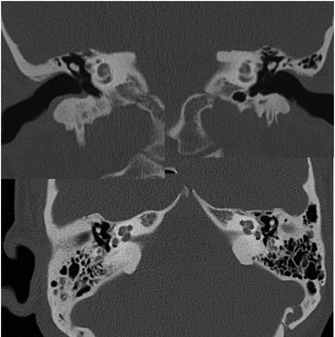
Selected axial and coronal temporal bone CT images demonstrate symmetric rounded zones of demineralization along the bilateral anterior oval window margins. There is superimposed mild inflammatory opacification of the right mastoid air cells with surrounding sclerosis. The right tympanic membrane is mildly thickened.
Discussion/Differential Diagnosis:
BACK TO
MAIN PAGE