Hemangiopericytoma
Findings:
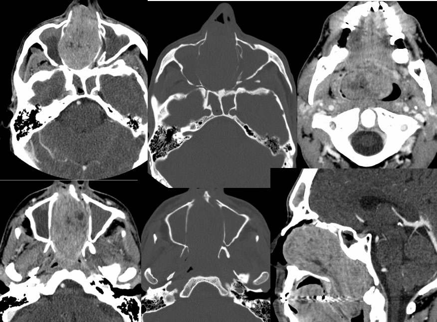
Multiple sinus CT images demonstrate a large enhancing mass involving the nasal cavity causing obstruction of the nasal cavity. The mass demonstrates subtle heterogeneity. There is adjacent bone remodeling. The mass extends posteriorly into the nasopharynx. There is superimposed low density inflammatory opacification of the maxillary and sphenoid sinuses potentially postobstructive.
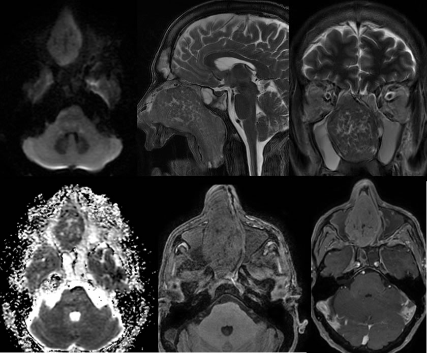
MR images also show nearly homogeneous enhancement of the mass, with relative decreased signal on T2 weighted imaging and evidence of diffusion restriction due to a cellular lesion. The postobstructive sinus inflammatory disease is confirmed to have different signal than the mass.
Discussion/Differential Diagnosis:
BACK TO
MAIN PAGE