Esthesioneuroblastoma
Findings:
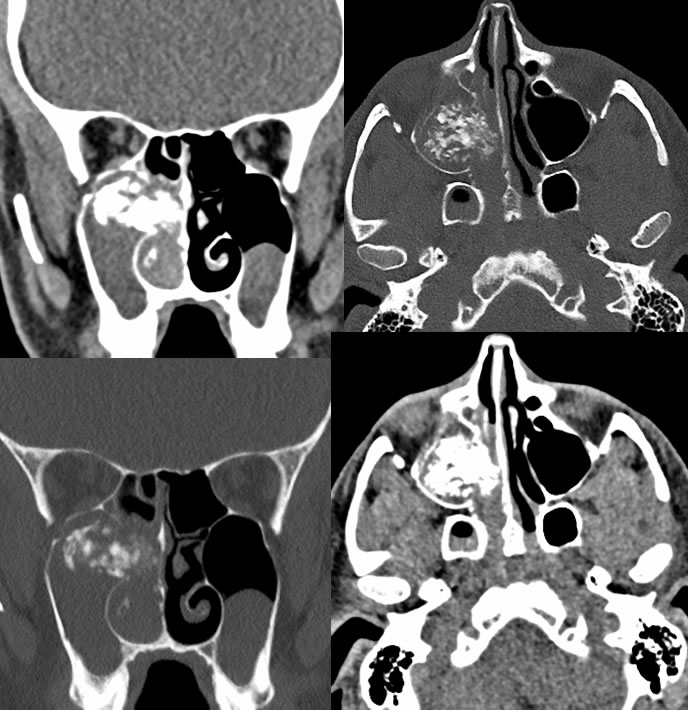
Noncontrast CT sinus images demonstrate a heavily calcified mass in the region of the right maxillary sinus ostium. There is postobstructive low-density opacification of the right maxillary sinus. The right nasal cavity is also obstructed and opacified with intermediate density soft tissue. A small to moderate mucus retention cyst is present in the left maxillary sinus. A portion of the right maxillary sinus medial wall is destroyed adjacent to the mass.
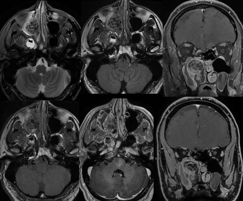
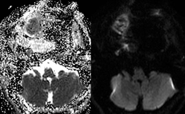
The MRI images demonstrate heterogeneous low level enhancement of the mass with the calcifications corresponding with zones of low signal. Diffusion weighted imaging and ADC map demonstrate probable mild diffusion restriction in the region of the mass. A superimposed capillary telangiectasia is present in the pons. Right sphenoid sinus inflammatory disease is present which has different signal than the mass. The postobstructive right
maxillary sinus inflammatory disease is again seen with uniformly enhancing peripheral mucosal thickening, again different than the signal of the mass.
Discussion/Differential Diagnosis:
BACK TO
MAIN PAGE