6 months later.
Next images are s/p Avastin therapy.
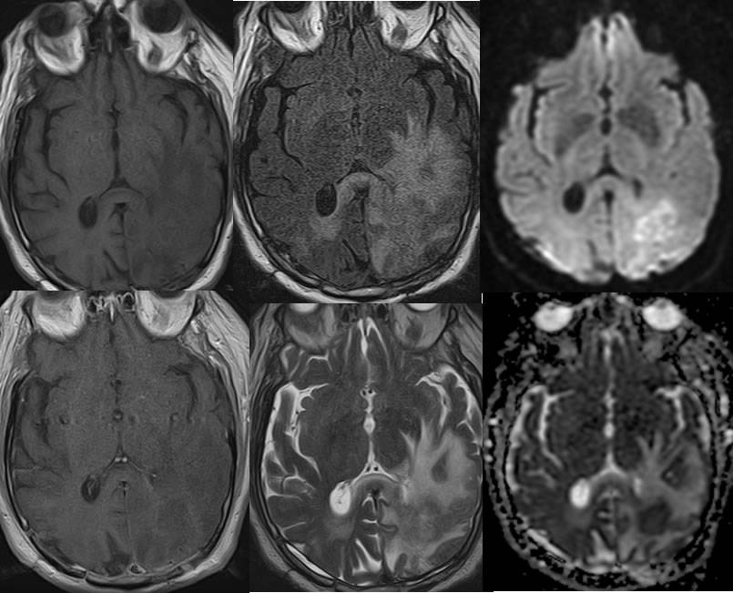
Recurrent GBM, s/p Avastin
Findings:
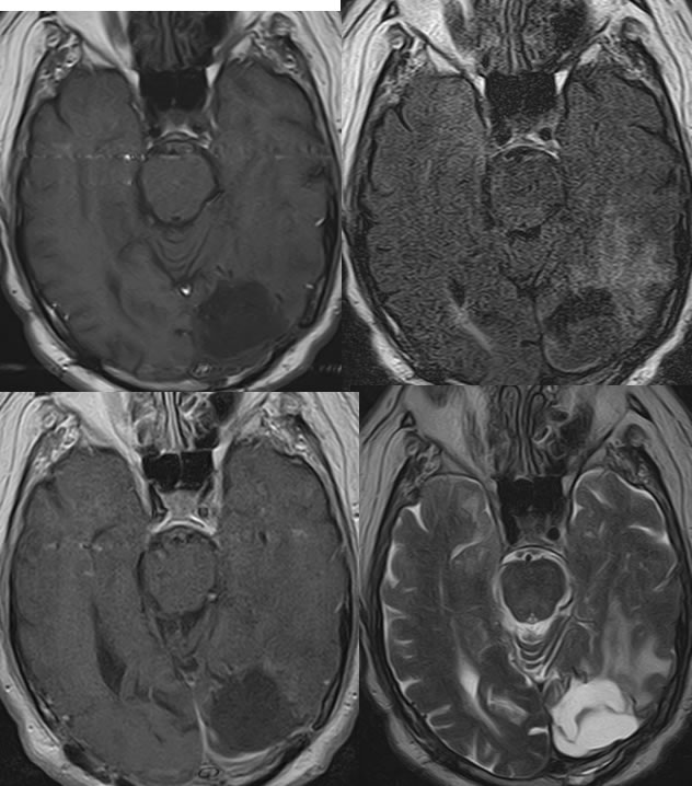
Baseline MRI images demonstrate postoperative findings of left parietooccipital craniotomy with an irregular surgical defect in the left occipital lobe. Radiation seeds line margins of the surgical defect with surrounding FLAIR signal alteration and no significant mass effect. Minimal poorly defined enhancement is present along the margins of the surgical defect. The surgical defect contains some hemorrhagic products.
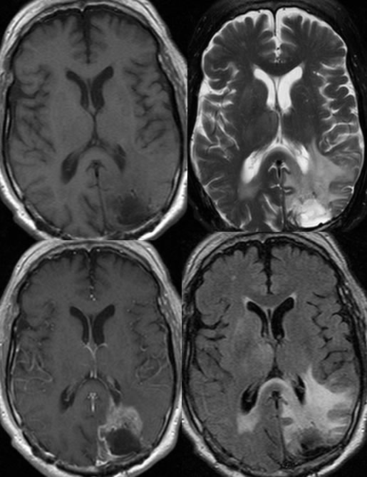
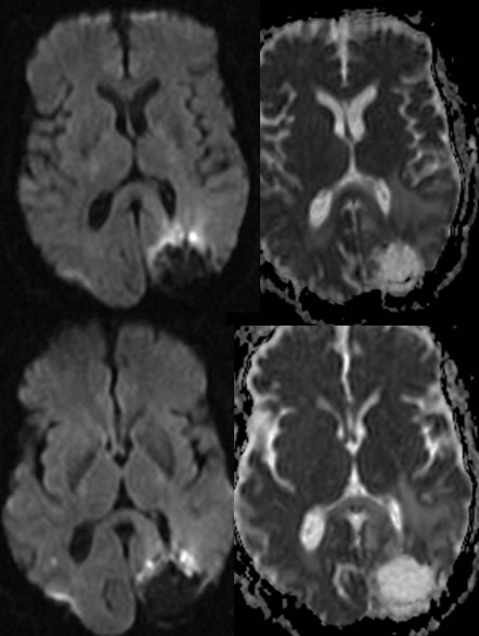
The follow up MRI six months later demonstrates significant progression of irregular enhancement along the margins of the surgical cavity with zones of diffusion restriction. The left occipital horn is partially effaced due to mass effect. The surrounding FLAIR signal alterations are progressive with extend into the corpus callosum splenium and right periatrial white matter.
Images after Avastin therapy demonstrate near resolution of the abnormal enhancement, however the mass effect, FLAIR signal alterations, and diffusion restriction have increased. The surgical defect has partially collapsed and contains more complex fluid.
Discussion/Differential Diagnosis:
BACK TO
MAIN PAGE