Recurrent GBM, dural dissemination
Findings:
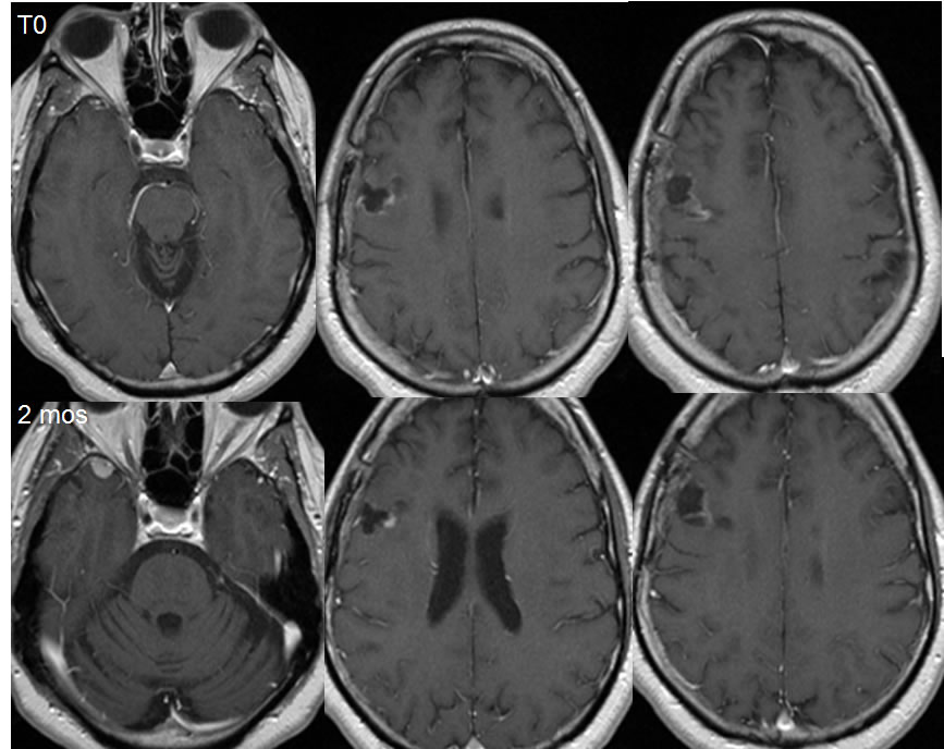
Baseline MRI post contrast images demonstrate postoperative findings of right parietal craniotomy with a small irregular surgical defect and mild associated enhancement. There is a subtle focus of extraaxial nodularity along the anterior right temporal lobe.
Two months later, the extraaxial mass over the right anterior temporal lobe has significantly increased in size. The zones of enhancement along the margins of the surgical defect are more evident.
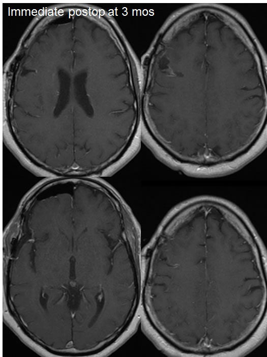
Immediate postoperative images demonstrate pneumocephalus and a small extraaxial fluid collection along the right cerebral convexity. The region of the right temporal extraaxial mass is not demonstrated on these images.
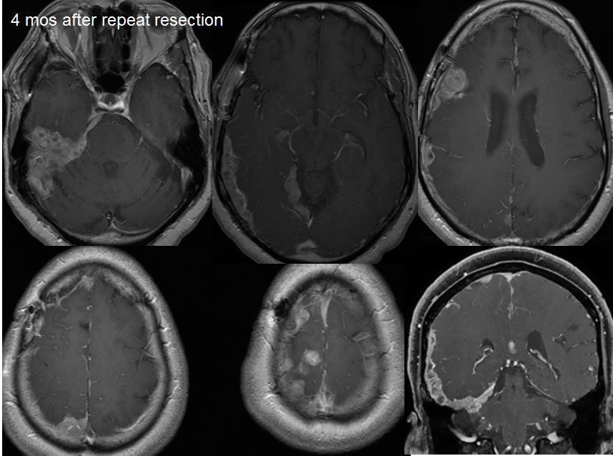
Four months after resection of the right anterior temporal extra axial mass, there is no definite local tumor recurrence with a small fluid collection in the region and mild surrounding linear enhancement. However, extensive nodular masses have developed along the dura of the right cerebral convexity due to extensive dural tumor dissemination.
Discussion/Differential Diagnosis:
BACK TO
MAIN PAGE