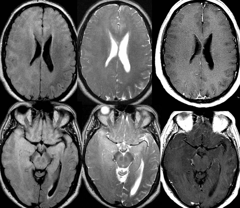
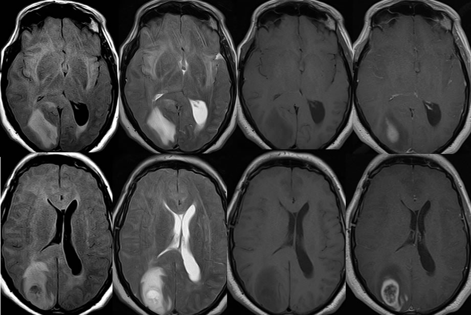
57 yo M, altered mental status and personality changes. Next images are 17 mos later.
Gliomatosis cerebri with GBM transfomation
Findings:
Initial MRI images demonstrate diffuse signal abnormality throughout both cerebral hemispheres right greater than left with mild mass effect on the right. These signal abnormalities also involve overlying cortex and are somewhat asymmetric in the medial temporal lobes.
17 months later, the diffuse signal alteration is slightly less evident, with slightly increased mass effect in the right cerebral hemisphere. A poorly defined peripherally enhancing mass has developed in the right parietal lobe with confluent surrounding FLAIR signal alteration, satellite zones of enhancement, and moderate localized mass effect. The left lateral ventricle has increased in size due to mild ventricular trapping.
Discussion/Differential Diagnosis:
BACK TO
MAIN PAGE