
Grade 3 Astrocytoma
Findings:
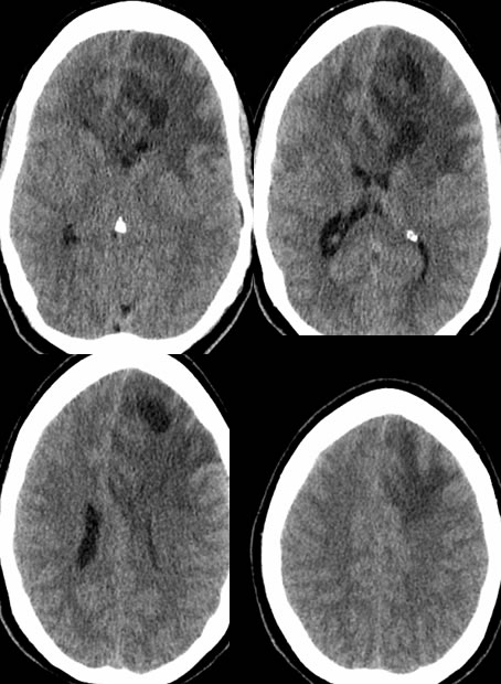
Noncontrast CT images demonstrate a mixed attenuation complex cystic mass in the bilateral frontal lobes left greater than right with localized mass effect and involvement of the anterior corpus callosum. Significant zones of hemorrhagic change are not confirmed. No calcification is visible.
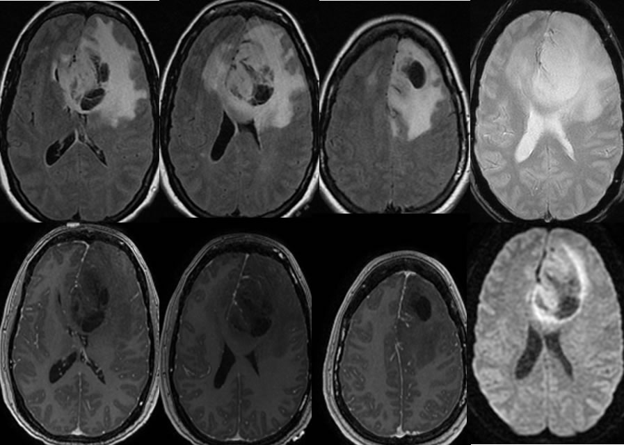
MRI images confirm a complex cystic overall nonenhancing mass with associated mass affect and surrounding FLAIR signal alteration. A few linear zones of enhancement are seen, in part likely vascular. Mixed diffusion characteristics are present with areas of hyperintensity.
Discussion/Differential Diagnosis:
BACK TO
MAIN PAGE

