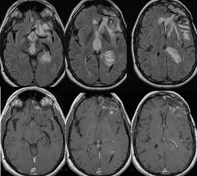
Images below are 18 months later.
Grade 3 anaplastic astrocytoma with intraventricular recurrence
Findings:
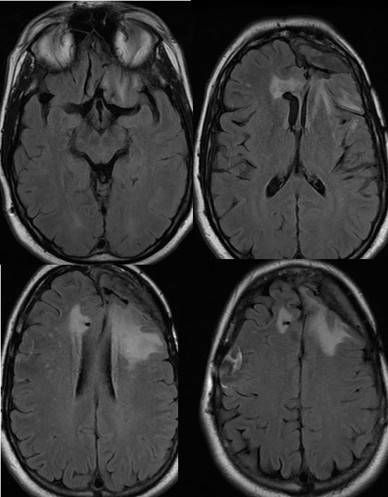
Axial FLAIR images demonstrate postoperative findings of left frontal craniotomy with an irregular surgical defect in the anterior left frontal lobe associated with tethering of the left frontal horn toward the operative defect. A shunt catheter enters from the right incompletely included with susceptibility artifact related to the reservoir over the right lateral frontal convexity. Patchy FLAIR signal alterations are seen within the bilateral frontal lobes left greater than right with no significant mass effect.
The follow up images 18 months later demonstrate development of multiple intraventricular masses with mild enhancement after gadolinium administration. There is additional enhancement and hemorrhagic change along the posterior margin of the surgical defect which is new. There is also infiltration of the hypothalamus and optic chiasm and proximal optic tract regions with the masses causing expansion of the ventricular system. Significant progression of signal alteration is seen within the anterior corpus callosum.
Discussion/Differential Diagnosis:
BACK TO
MAIN PAGE