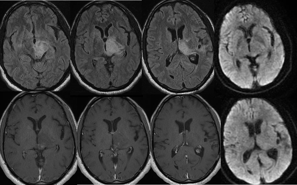
3 months after biopsy below, nonspecific inflammatory with no evidence of neoplasm.
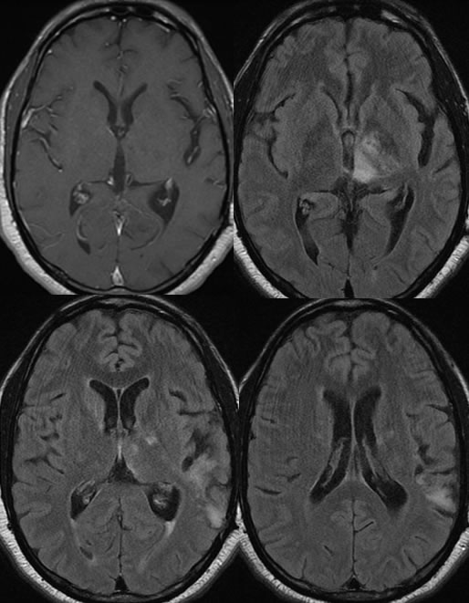
3 month follow up below, after steroid therapy.
Encephalitis, unknown etiology, possible IRIS
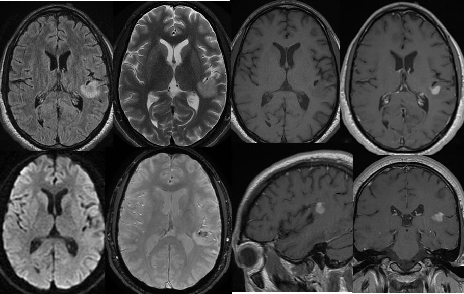
Findings:
The initial MRI images demonstrate a focus of irregular enhancement posterior to the left insula with surrounding T2 hyperintensity, minimal hemorrhagic staining, and no diffusion restriction. There is subtle localized mass effect.
The follow up images after biopsy demonstrate a dramatic improvement of signal alteration and resolution of enhancement along the posterior right insula, but there is now a larger zone of patchy hyperintensity involving the left thalamus. The new lesion is associated with a few patches zones of enhancement along the posterior aspect, again with no diffusion restriction. There is localized mass effect with partial effacement of the third ventricle.
The follow up images after steroid therapy demonstrate improvement of signal alteration in the left thalamus with resolution of abnormal enhancement. A new focus of developing signal alteration is present in the left parietal lobe.
Discussion/Differential Diagnosis:
BACK TO
MAIN PAGE