Chondrosarcoma
Findings:
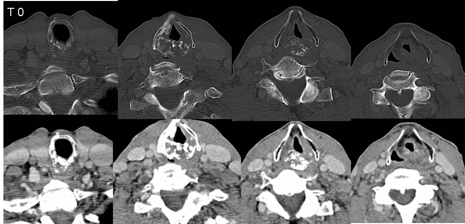
Initial CT images demonstrate and expansile lytic lesion involving the cricoid cartilage with stippled zones of calcified internal matrix. Irregular calcification in the right paraglottic region may be related to previous augmentation.
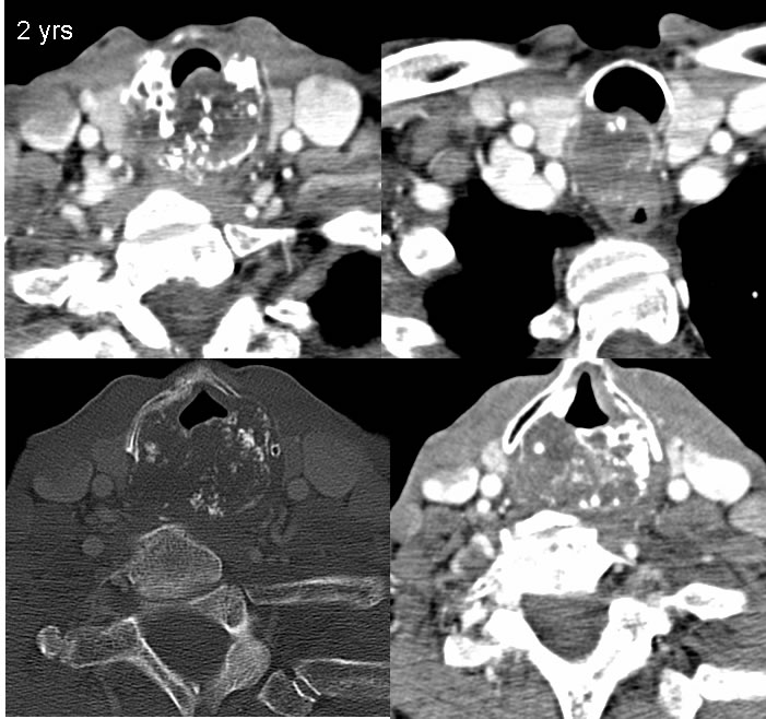
The follow up images two years later demonstrate marked interval expansion of the mass, with more soft tissue components and more infiltrative characteristics. The mass causes moderate effacement of the tracheal air column. The mass is also inseparable from the ventral margin of the proximal esophagus. The mass infiltrates the posterior wall of the trachea.
Discussion/Differential Diagnosis:
BACK TO
MAIN PAGE