Spinocerebellar Ataxia Type 3
Findings:
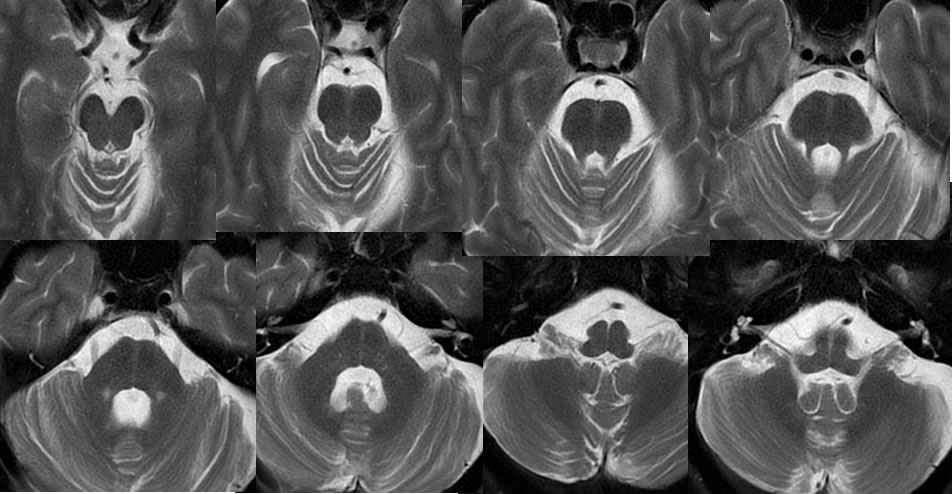
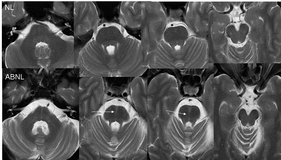
Multiple T2 weighted MRI axial images centered on the pons and cerebellum demonstrate atrophy of the midbrain and pons, associated with a sagittally oriented band of hyperintensity coursing from anterior posterior through the pons, with volume loss in this region. Mild cerebellar atrophy is incompletely included.
With a normal patient for a comparison, the hyperintense band is not seen and the volume loss of the midbrain is not evident. No significant cerebellar atrophy is visible in the control patient.
Discussion/Differential Diagnosis:
BACK TO
MAIN PAGE