Thyroid Lymphoma
Findings:
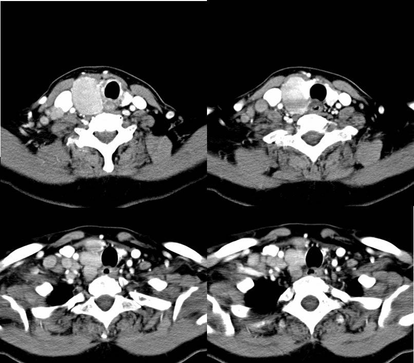
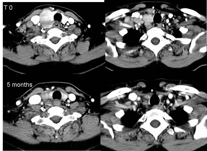
Axial contrast enhanced neck CT images demonstrate a homogeneous and diffusely low level enhancing mass involving the right thyroid lobe with normal appearance of the included left thyroid lobe. There is associated right supraclavicular adenopathy which does not appear necrotic. The mass enhances less than normal thyroid tissue.
Five months later without evidence of surgery, the mass has dramatically decreased in size and the adenopathy is no longer visible.
Discussion/Differential Diagnosis:
BACK TO
MAIN PAGE