Leptomeningeal Carcinomatosis, Breast. Superimposed L sigmoid sinus thrombosis.
Findings:
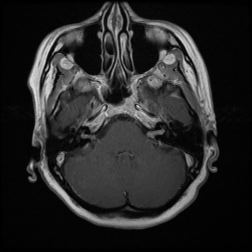
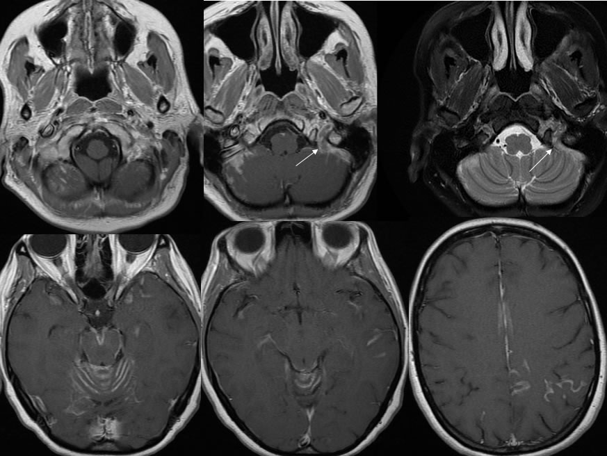
The initial post contrast axial T-1 weighted image demonstrates relatively symmetric abnormal enhancement within both internal auditory canals. The subsequent images demonstrate abnormal leptomeningeal enhancement coating the brainstem and cerebellar vermis, with several other spotty areas of leptomeningeal enhancement bilaterally within cerebral sulci and along the right cerebellum. The abnormality in the right cerebellum demonstrates T2 hyperintensity with no significant surrounding edema. The arrows demarcate a zone of thrombosis of the left sigmoid sinus with T2 hyperintensity and decreased enhancement within the lumen of the left sigmoid sinus.
Discussion/Differential Diagnosis:
BACK TO
MAIN PAGE