Epidural and Leptomeningeal Metastases, Lung Cancer
Findings:
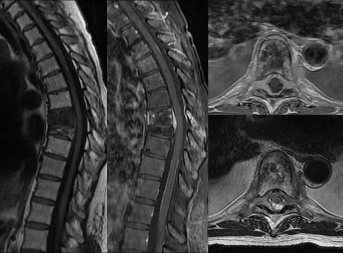
The sagittal T1 weighted image demonstrates abnormal hypointensity of the T7 vertebral body with collapse of the T8 vertebral body. The T8 vertebral body has spotty generally fatty marrow and there is marrow replacement of the T7 vertebral body which also involves posterior elements. The sagittal fat saturated T1 weighted image demonstrates abnormal enhancement in the ventral and dorsal epidural space at T7. There is also abnormal linear irregular enhancement coating the thoracic cord most prominent distally, and also seen dorsally at the T3-4 level. The axial post contrast image demonstrates abnormal nearly circumferential epidural enhancement at the T7 level causing relative effacement of CSF around the cord. There is no significant cord compression. The abnormal enhancement extends into the paravertebral regions left slightly greater than right and within both foramina. Bilateral pleural effusions are incompletely included.
Discussion/Differential Diagnosis:
BACK TO
MAIN PAGE