Grade 2 Neurocytoma, Pineal region
Findings:
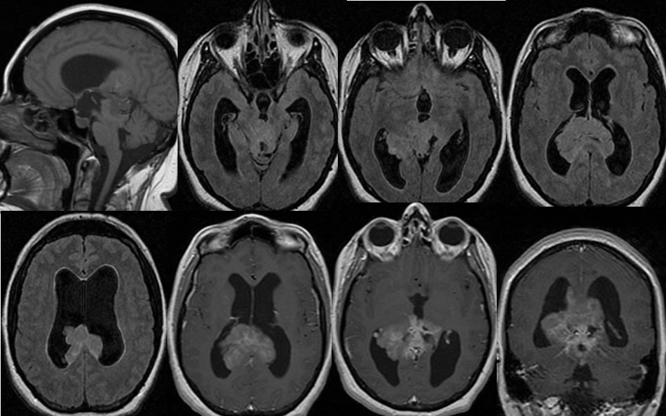
Multiple MR images demonstrate a poorly defined heterogeneously enhancing infiltrative mass at midline invading the bilateral ventricular system, dorsal midbrain, and supravermian cistern. The mass is only slightly hyperintense to normal brain on the FLAIR images. The lateral ventricles are dilated due to obstructive hydrocephalus at the level of the cerebral aqueduct. On the sagittal T1 image, the infiltrative mass is localized to the expected region of the pineal gland. Normal pineal gland is not visible.
Discussion/Differential Diagnosis:
BACK TO
MAIN PAGE