Gliosarcoma
Findings:
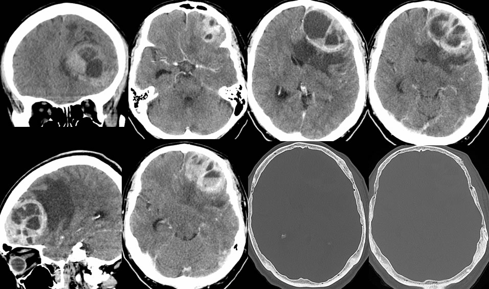
Multiple CT images demonstrate a heterogeneously enhancing intraaxial mass in the left frontal lobe with significant surrounding vasogenic edema and midline shift of the septum pellucidum. The mass contains fluid fluid levels likely due to hemorrhagic components within complex cystic foci. A focus of nodular subgaleal enhancement and cortical irregularity of the outer table indicates transosseous extent of the mass into the subgaleal space. The mass is faintly hyperdense precontrast due to cellularity.
Discussion/Differential Diagnosis:
BACK TO
MAIN PAGE