
Epidermoid
Findings:
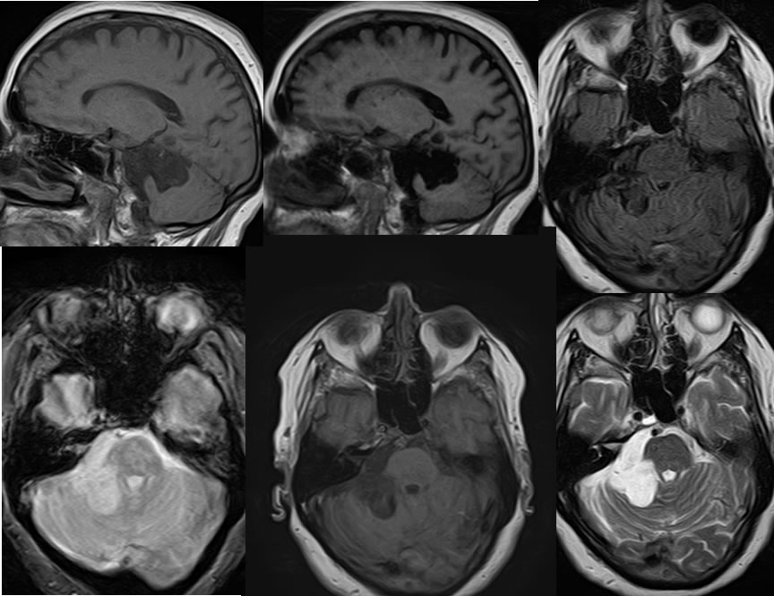
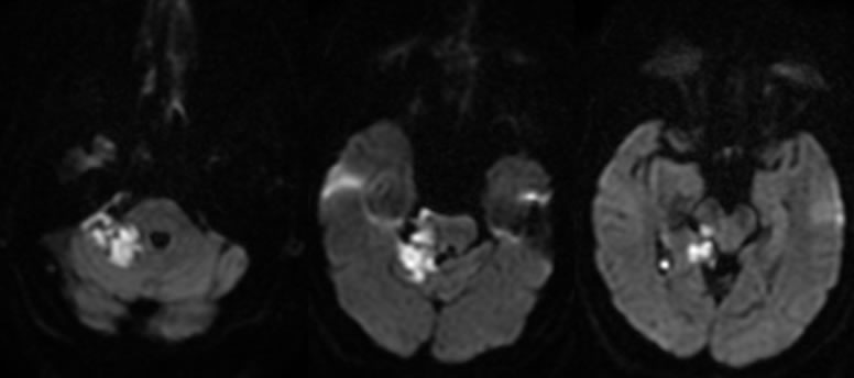
Multiple MRI images demonstrate a multi lobulated near CSF signal mass with in the right CP angle cistern causing mass effect on the cerebellum and brachium pontis. The mass shows subtle hyperintensity to CSF on the T1 weighted imaging and does not follow CSF signal on FLAIR imaging. No hemorrhagic change is seen in the region. On diffusion weighted imaging, the mass has heterogeneous patchy zones of hyperintensity due to restricted diffusion.
Discussion/Differential Diagnosis:
BACK TO
MAIN PAGE

