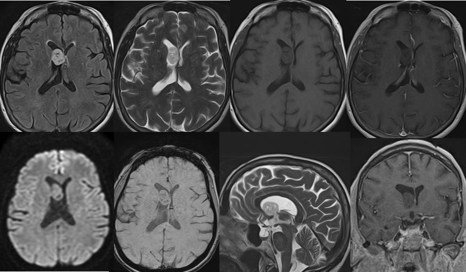
Subependymoma
Findings:
Multiple MRI images demonstrate a well-defined ovoid nonenhancing mass with in the frontal horn of the right lateral ventricle. The mass demonstrates T2 hyperintensity, T2 hypointensity, and no hemorrhagic change. There is mild asymmetric dilation of the right lateral ventricle. Spotty zones of low signal within the mass may reflect calcifications.
Discussion/Differential Diagnosis:
BACK TO
MAIN PAGE