Grade 2 Astrocytoma
Findings:
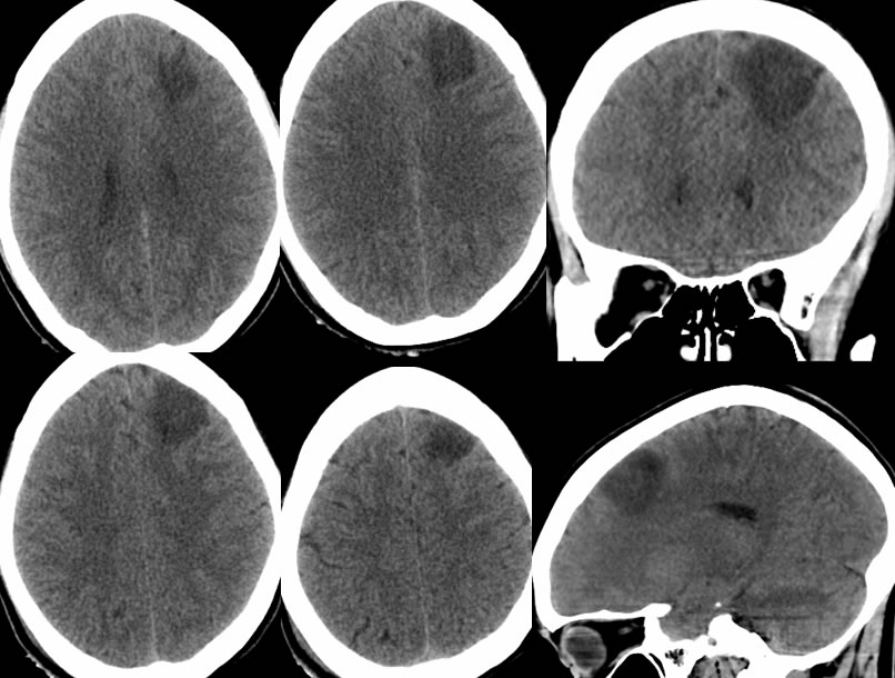
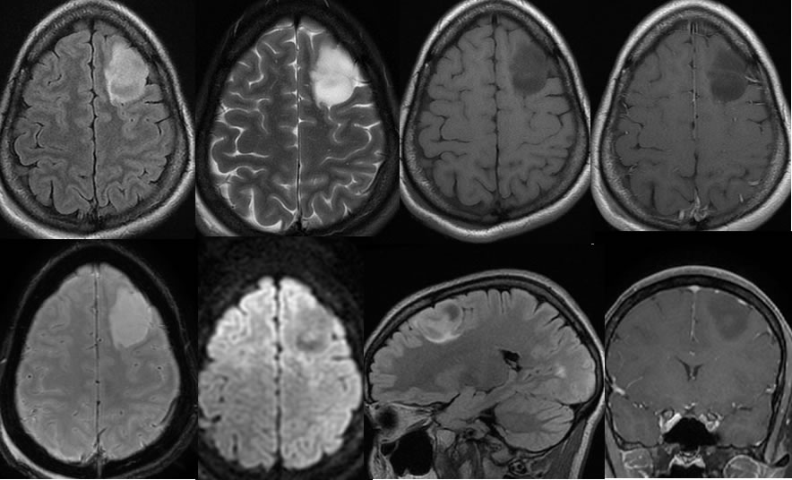
Noncontrast CT images demonstrate a somewhat wedge shaped focus of low attenuation in the left frontal lobe with mild expansile characteristics suggested. The low attenuation is well defined. On the MRI images, a nonenhancing expansile mass is visible in this region which causes marked expansion of the middle frontal gyrus. Minimal FLAIR hyperintensity is seen along the margins of the lesion inferiorly.
Discussion/Differential Diagnosis:
BACK TO
MAIN PAGE