Subependymoma
Findings:
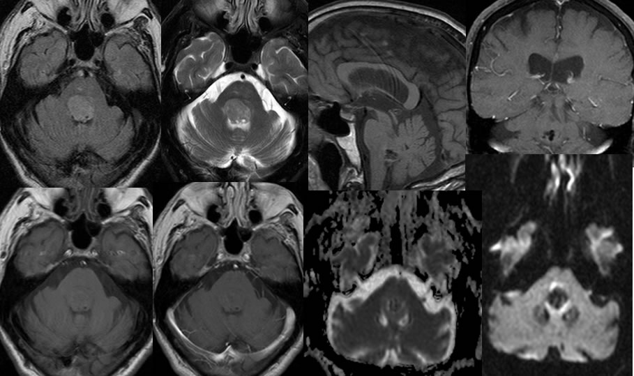
Multiple MRI images demonstrate a well circumscribed nonenhancing mass without restricted
diffusion within the fourth ventricle. Zones of low signal in the inferior margin of the mass are most compatible with calcifications. The mass is near isointense on T1 weighted imaging and minimally hyperintense on FLAIR and T2. There is no significant surrounding vasogenic edema. The fourth ventricle is slightly effaced but no significant hydrocephalus is included.
Discussion/Differential Diagnosis:
BACK TO
MAIN PAGE